


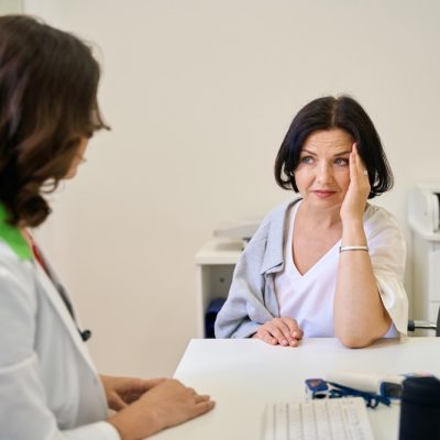
Diagnosis
AI-powered mammograms: a new window into heart health

Mammograms used in combination with AI may reveal much more than cancer, but can also be used to assess the amount of calcium build up in the arteries within breast tissue – an indicator of cardiovascular health, a new study shows.
While breast artery calcifications can be seen on mammogram images, radiologists do not typically quantify or report this information to women or their clinicians.
This new study, which used an AI image analysis technique not previously used on mammograms, demonstrates how AI can help fill this gap by automatically analysing breast arterial calcification and translating the results into a cardiovascular risk score.
“We see an opportunity for women to get screened for cancer and also additionally get a cardiovascular screen from their mammograms,” said Theo Dapamede, postdoctoral fellow at Emory University in Atlanta and the study’s lead author.

“Our study showed that breast arterial calcification is a good predictor for cardiovascular disease, especially in patients younger than age 60. If we are able to screen and identify these patients early, we can refer them to a cardiologist for further risk assessment.”
Heart disease is the leading cause of death in the United States but remains underdiagnosed in women and there is also lagging awareness. Researchers said the use of AI-enabled mammogram screening tools could help identify more women with early signs of cardiovascular disease by taking better advantage of screening tests that many women routinely receive.
A build up of calcium in blood vessels is a sign of cardiovascular damage associated with early-stage heart disease or aging. Previous studies have shown that women with calcium build up in the arteries face a 51 per cent higher risk of heart disease and stroke.
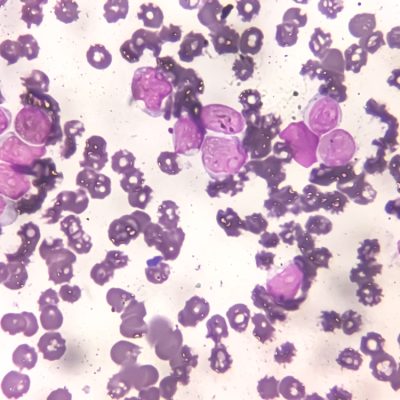
To develop the screening tool used for this study, researchers trained a deep-learning AI model to segment calcified vessels in mammogram images, which appear as bright pixels on X-rays, and calculate the future risk of cardiovascular events based on data obtained from the electronic health record data.
The segmentation approach is what separates this model from previous AI models developed for analysing breast artery calcifications. Researchers said the model is also strengthened by its use of a large dataset for training and testing, which included images and health records from over 56,000 patients who had a mammogram at Emory Healthcare between 2013 and 2020 and had at least five years of follow-up electronic health records data.
“Advances in deep learning and AI have made it much more feasible to extract and use more information from images to inform opportunistic screening,” Dapamede said.
Overall findings showed the new model performed well at characterizing patients’ cardiovascular risk as low, moderate or severe based on mammogram images.
After calculating the risk of dying from any cause or suffering an acute heart attack, stroke or heart failure at two years and five years, the model showed that the rate of these serious cardiovascular events increased with breast arterial calcification level in two of the three age categories assessed – women younger than age 60 and age 60 to 80, but not in those over age 80.
This makes the tool particularly well suited for providing early warning of heart disease risk in younger women, who can benefit more from early interventions, researchers said.
The results also showed that women with the highest level of breast arterial calcification (above 40 mm2) had a significantly lower five-year rate of event-free survival than those with the lowest level (below 10 mm2).
For example, 86.4 per cent of those with the highest breast arterial calcification survived for five years compared with 95.3 per cent of those with the lowest level of calcification. This translates to approximately 2.8 times the risk of death within five years in patients with severe breast arterial calcification compared to those with little to no breast arterial calcification.
The AI model was developed as a collaboration between Emory Healthcare and Mayo Clinic and is not currently available for use.
If it passes external validation and gains approval from the U.S. Food and Drug Administration, researchers said the tool could be made commercially available for other health care systems to incorporate into routine mammogram processing and follow-up care.
The researchers also plan to explore how similar AI models could be used for assessing biomarkers for other conditions, such as peripheral artery disease and kidney disease, that might be extracted from mammograms.


A new reporting tool built specifically for obstetrics and maternal-fetal medicine has launched, aimed at teams managing increasingly complex pregnancies with limited time and resources.
Trice Imaging has released Trice Workspace Reporting, which connects imaging, reporting and longitudinal patient data in a single workflow to support faster clinical decision making.
Birth rates are falling worldwide, but pregnancies are getting more complicated. Advanced maternal age, IVF-assisted pregnancies, rising obesity rates and a higher prevalence of hypertension and diabetes mean more cases now require specialist monitoring, advanced imaging and multidisciplinary care.
At the same time, clinical teams are stretched and facing growing administrative demands.
Trice Workspace Reporting brings together customisable reporting, dynamic pregnancy dating and longitudinal patient history with an AI-ready, EHR-interoperable infrastructure, all inside the company’s Tricefy image management platform.
The company says it aims to accelerate standardised and synchronised report turnaround, support timely clinical decisions and improve operational efficiency for fetal medicine services.
“Maternal fetal medicine teams are managing increasingly complex pregnancies while being asked to do more with limited time and resources,” said Mark A. Samii, chief revenue officer at Trice Imaging.
“Trice Workspace Reporting is designed to remove unnecessary friction from reporting by creating a structured digital foundation that supports today’s need for connected clinical workflows.
“It also provides a digital foundation as practices prepare for tomorrow and the evolution of AI-enabled fetal assessment, anomaly detection and outcome prediction technologies.”
Trice Imaging describes its mission as transforming the women’s health journey by connecting physicians, patients and healthcare systems. From independent practices to large hospital ecosystems, it aims to reach the entire women’s health continuum, spanning IVF and reproductive health, maternal-fetal medicine and OB/GYN, and onwards to lifelong women’s health.
For more than 17 years the firm has worked on cloud-based storage, retrieval, display, organisation and exchange of ultrasound medical images and associated information across health environments.
Its wider platform now extends to dynamic clinical reporting, AI-driven workflow optimisation, data analytics and secure patient engagement.
Trice Imaging holds regulatory and data protection clearances in 40 countries. It has offices in Miami and Stockholm, alongside a growing network of global distributors.

Two endometriosis tests could cut years from diagnosis after NICE backed their temporary NHS use in England and Wales.
EndoSure and Endotest have been recommended in draft guidance, with one able to provide results in 45 minutes.
Endometriosis affects around one in 10 women of reproductive age. It occurs when tissue similar to the womb lining grows elsewhere, including around the ovaries and fallopian tubes.

Symptoms can include painful periods, painful bowel movements, pain when urinating and pain during or after sex.
Diagnosis can involve ultrasound scans, magnetic resonance imaging (MRI) or laparoscopy. A laparoscopy is keyhole surgery in which a camera is inserted through a small cut in the abdomen.
Despite the effect the condition can have on physical and mental health, women can wait years for a diagnosis.
The average wait in the UK is nine years and four months, rising to 11 years for women from ethnically diverse communities, according to the National Institute for Health and Care Excellence (NICE).
Long waits can increase suffering, prolong poor health and allow the condition to progress, making it more difficult to treat.
Dr Anastasia Chalkidou, NICE’s healthtech programme director, said: “A diagnosis of endometriosis can for some women take the best part of a decade, with the UK average standing at nine years and four months, and rising to 11 years for those from ethnically diverse communities.”
She said delays could lead to chronic pain affecting daily life, relationships and work.
She added: “These technologies have the potential to change that by giving primary care professionals better non-invasive tools to identify endometriosis earlier, allowing earlier and better treatment.
“Our draft guidance reflects our commitment to getting promising innovations to patients quickly, while making sure the evidence to support their wider use is built in a rigorous way.”
Endotest examines a saliva sample for microRNAs, tiny biological markers that can indicate the presence of endometriosis.
The sample is sent to a laboratory and the result returned to a GP or another healthcare professional to inform the next steps in diagnosis and care.
EndoSure uses sensor pads placed on the abdomen to measure electrical signals in the gut.
Women must fast for between six and eight hours before the 45-minute test. During the procedure, they drink water until they feel full, helping the device record gut activity accurately.
Results are available as soon as the test is complete.
The draft recommendation, published on Tuesday, approves both technologies for three years while further evidence is collected on how well they work.
NICE will then decide whether to approve them permanently for NHS use.
NICE said a third test, DotEndo, needs more research before it can be recommended.
EndoSure and Endotest are not designed to diagnose the condition on their own.
They are intended for women whose symptoms still suggest endometriosis after a normal clinical examination and negative or inconclusive imaging results, or when imaging has not been carried out.
Dr Gail Busby, a consultant gynaecologist at Manchester University NHS Foundation Trust, said: “These tests are a gamechanger because they give us answers much earlier, without the need for invasive surgery, and that means we can start the right treatment sooner.
“An earlier diagnosis doesn’t just change one person’s life, it frees up appointments and surgical slots for everyone waiting for care.”
Emma Cox, of Endometriosis UK, welcomed the tests.
She said their introduction should be supported by education for GPs and nurses to ensure prompt access and prevent symptoms from going unrecognised.

Xella Health has launched what it calls the first AI precision health platform built for the XX chromosome.
The company says it aims to address a lack of diagnostic precision and clinical research focused on female biology.
Women make up half of the population and account for 80 per cent of consumer healthcare decisions, but research into women’s health has historically received less funding than male-focused studies.

Kelly Lacob, Xella Health co-founder and chief executive, said: “Women have been trapped in a diagnostic dark age experiencing debilitating symptoms like severe period pain, bloating and GI issues, exhaustion, and brain fog, routinely dismissed by the healthcare system.
“This dismissal results in women being diagnosed four years later than men, on average, for the same conditions, and a seven-to-10-year delay for women to receive an accurate diagnosis for conditions like endometriosis.
Stalling necessary care and treatment results in prolonged suffering with chronic pain, heightened infertility risks, and declining mental health.
Xella is here to replace the systemic medical gaslighting women have endured for generations.
We are handing women the evidence and information they need to advocate for themselves and secure faster, accurate diagnoses before early-stage conditions spiral.”
Xella says its AI examines billions of data points from clinical information and multi-omic biomarkers to assess the probability of more than 130 conditions specific to female biology.
Multi-omic data combines information from several biological areas, including genes, proteins and hormones.
The conditions assessed include polyendocrine metabolic ovarian syndrome, or PMOS, formerly known as polycystic ovary syndrome, as well as perimenopause and endometriosis.
Xella was founded by Lacob, Adriana Dantas and Dr Jesus Ching, who developed the concept while working together on molecular diagnostics at Mammoth Biosciences.
The founders say the platform is designed to provide information about possible underlying causes through advanced testing and long-term care of a kind often available only through expensive concierge services.
They drew on personal experiences to build a service intended to identify small changes in a woman’s biological baseline.
Members complete an initial health questionnaire before having blood taken at a local partner laboratory such as Quest or Labcorp.
A phlebotomist can also visit a member’s home for an additional charge.
The company’s AI analyses biomarker data from genomics, proteins and hormones alongside symptoms, lifestyle risks and medical history.
Xella says this information is used to screen for more than 130 female-specific conditions, including PMOS, Hashimoto’s disease, premenstrual dysphoric disorder, endometriosis and perimenopause timelines.
Hashimoto’s disease is an autoimmune condition in which the immune system attacks the thyroid gland.
Premenstrual dysphoric disorder, or PMDD, is a severe form of premenstrual syndrome that can cause significant emotional and physical symptoms.
The results are processed through Xella’s own dry laboratory, which the company says is certified under the US Clinical Laboratory Improvement Amendments and accredited by the College of American Pathologists.
A dry laboratory analyses data using computing and other non-experimental methods rather than carrying out traditional laboratory procedures.
The findings are turned into a personalised healthcare plan and reviewed with a certified telehealth doctor.
The doctor may recommend immediate clinical action, including personalised hormone therapy or referrals to genetic counsellors, pelvic floor physiotherapists and reproductive endocrinologists.
Reproductive endocrinologists are doctors who specialise in hormones, fertility and reproductive health conditions.
Dantas, co-founder and chief operating officer, said: “Women’s health data has historically been treated in isolated silos – a hormone test here, an ultrasound there – but no one was connecting the dots across the entire biology.
“By tracking unique biological patterns longitudinally across cycles and life stages, we aren’t just providing data, but a clear path forward.”
Xella’s clinical advisers include Dr Allison Kurian, director of Stanford Women’s Clinical Cancer Genetics Program and professor of medicine, epidemiology and population health at Stanford.
They also include Dr Lynn Westphal, a reproductive endocrinology and infertility specialist and chief medical officer of Kindbody.
Xella has received US$4.7m in angel and pre-seed funding from Precursor Ventures, Capital F, Ulu Ventures and Swizzle Ventures.
Other funds and angel investors from healthcare, diagnostics and consumer technology also participated.
Margaret Coblentz, co-founder and general partner of Capital F, said: “Women’s health is one of the highest-momentum categories in the market today, driven by a US$15tn female economy.
“Xella represents exactly how Capital F sees women’s health evolving: deep clinical expertise paired with a consumer-first mindset, and a genuine opportunity to unlock the next generation of healthcare.”


 Menopause1 week ago
New menopause drug approved for use by NHS in Scotland

 Entrepreneur2 weeks ago
Applications open for the third W Accelerate with Merck KGaA and M Ventures

 Hormonal health7 days ago
Stardust period tracker shares health data, study reveals

 News2 days ago
Zero Candida market set to reach over US$2 billion by 2030 – report

 News1 week ago
Women’s health draws record $1.55bn in equity as capital spreads beyond the mega-rounds

 Mental health2 weeks ago
SSRIs may lower heat intolerance in women with depression – study

 News2 weeks ago
Avni Wellness secures US$470k funding

 Wellness2 weeks ago
Alcohol and smoking linked to breast cancer and irregular heartbeat in women, study finds