




News
Melinda French Gates-backed startup Tia cuts 23% of staff


Women’s healthcare company Tia, backed by Melinda French Gates, has cut 72 jobs, reducing its workforce by 23 per cent across corporate and clinical divisions.
Chief executive Felicity Yost told staff on Monday that the company would downsize as part of a plan to accelerate its path to profitability, according to Business Insider.
The cuts affected 17 corporate employees, 27 clinical providers and 28 field support operations roles.

Yost said feedback received during recent fundraising discussions prompted the company’s reassessment.
In an internal memo reported by Business Insider, she said Tia had been told to “rethink our business in the current economic and policy climate, which is one that prizes cost and profit-consciousness.”
A Tia spokesperson said the company “has seen strong growth, particularly in membership, which has outpaced our expectations for 2025.”
Beyond the job cuts, Tia has reduced executive pay and is reviewing vendor contracts to lower costs.
Management told employees they need to “creatively rethink how we optimise our administrative and care support roles across a broader range of responsibilities.”
Tia operates 11 clinics across the US — six in Los Angeles, two in New York City, one in San Francisco and two in Arizona.
The company provides integrated healthcare for women, including primary care, gynaecology, mental health support, and complementary treatments such as acupuncture and skincare.
Patients can choose between two payment models: Tia Essential, which allows pay-per-visit access using insurance, and a US$25-per-month membership that offers expanded benefits such as priority scheduling and coordinated lab and referral support.
The startup raised US$100m in a Series B funding round in 2021 and later secured extension funding from Pivotal Ventures, founded by Melinda French Gates, bringing total investment to US$150m.











Manchester City has launched a female athlete health platform covering menstrual, pelvic and breast health, as well as nutrition.
The Her City website is designed for the club’s women’s first-team players, coaches, support staff, academy players and parents.
Manchester City says the platform is the first of its kind in the Women’s Super League and was developed over two years.
The project was led by director of performance services Emma Deakin, physical performance scientist Rosie Anderson and PhD student and first-team nutritionist Sarah Malone.
It grew out of PhD research into menstrual, pelvic and breast health and nutrition, with the team seeking to turn that work into an online educational resource.
The website divides the four areas into separate sections and provides peer-reviewed information that the club says has been critically analysed by experts.
Content will continue to be reviewed and updated as further evidence emerges.
Resources include posters, visual explainers, audio materials and downloadable educational content, with information tailored to different groups including players and parents.
The platform aims to improve knowledge, develop critical analysis skills and strengthen communication within football and at home.
It also includes material to help users assess misinformation they encounter on social media or elsewhere.
A confidential contact form allows users to raise concerns or ask questions directly of experts at Manchester City.
Parents of academy players can also access the platform, with resources intended to help them support their children during a key phase of their development.
Manchester City said its longer-term aim is to make the platform available to all women working across City Football Group.


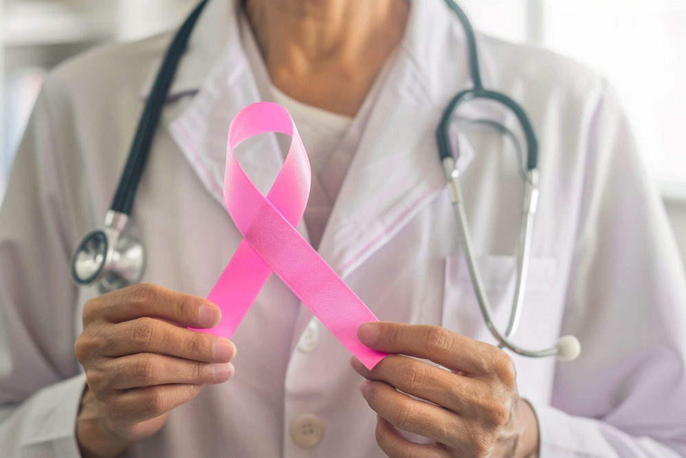
Winner of the Women’s Cancer Innovation Award at the 2026 Femtech World Awards, Namida Lab is working to tackle gaps in breast cancer screening and detection through the development of an innovative tear-based test called Aria.
Catching breast cancer early depends on accessible diagnostics, with research showing that geographic inaccessibility is the most significant barrier to early detection and diagnosis.
Equally, surveys show that almost 50 per cent of US women who are eligible for an annual mammogram do not receive one every year.
Namida Lab is working to address these gaps with its breast cancer screening test, Auria.
By identifying biomarkers in tears, the-home test offers a cost-effective, accessible way that aims to improve access and uptake.
The test does not diagnose breast cancer, but detects signals early on that indicate breast cancer may be present.
Omid Mogadam, CEO of Namida Lab, speaks to Femtech World about how the company aims to save lives by improving early detection, and what it means to win the Femtech World Women’s Cancer Innovation Award 2026.
The Auria test has a unique way of detecting breast cancer using tears – what was it that inspired you to use tears as a way of detecting breast cancer?
Our work comes from academic research that is around 20 years old. There were a number of breast cancer surgeons at the forefront of trying to find early screening, because they were the ones who had to deal with consequences of finding cancers in later stages.
Two of these surgeons we know: Suzanne Love at UCLA and Suzanne Klimberg at UAMS.
They started looking at biomarkers in alternate fluids other than blood, and Suzanne Love discovered cancer or breast cancer markers in milk of lactating women – nickel aspirate.
Klimberg started looking at tears because the nipple aspirate and tears are both byproducts of blood plasma.
They did clinical trials and found actually that there was a difference between the protein levels in tears of women with and without breast cancer.
That was the basis of the work that we adopted and brought into the company; to actually identify what those markers were, and to validate them through various trials, and then turn that into the product that eventually became Auria.
What makes tears unique is that there’s a lot of dead cells and pieces of other analytes that are circulating in blood.
They are much larger proteins which mask the smaller ones that you’re looking for. These cancer markers are typically small small molecules, and finding them in blood becomes an expensive proposition.
What are the gaps in diagnostic care that need addressing?
Our modern healthcare system is very good at advanced diagnostics in treatments, new treatments, and advanced imaging.
What it’s not good at is engagement, in bringing people in at an early stage.
In order to be able to serve everyone, keep people healthy, and not bankrupt the healthcare systems, you really do need that early engagement, which currently doesn’t exist.
A test like ours uses a signal from your body to tell you that you need to engage the system, and that is very powerful.
The result of our test is not whether you have breast cancer, it says that there is a signal that says there might be breast cancer – so, you need to follow up and engage sophisticated imaging, diagnostics, and treatment in the healthcare system.
As a result, more people will screen, and we will find cancers in earlier stages.
Right now, in the U.S. half the mortality in breast cancer is in women under the age of 45, and a lot of them have never been screened. They come in with later stage cancers and we need to flip that statistic.
Our test is recommended for someone without symptoms, and who may not be a high risk person. If you’re high risk, you need to be in a high risk screening programme, but this is for people of average risk with no symptoms.
Can you explain the science behind how the screening test works with proteins in tears to detect the possibility of breast cancer being present?
Looking at the early cancer detection technologies, there are a lot of products that use circulating tumor DNA and methylated DNA.
These all all fall under the same category of DNA tests, and they look for the DNA shedded cells from tumours.
There is a negative to using ctDNA or methylated DNA for early cancer detection because, in early cancer detection, there’s not enough of those shed cells because the tumor has not formed or has formed it very small and it’s not shedding.
This means that these types of tests do very well in later stages of cancer.
For earlier stages, you shouldn’t be looking for DNA. That’s why we focus on proteins.
We’re looking for proteins that surround the formation of cancer. In breast we’re looking for breast inflammation, and vascularization proteins, which always exist in the body.
So, those are the proteins that we’re looking for, and we’re looking for elevation of those proteins. We have had several rounds of discovery in order to identify those proteins.
The very first one, we took human tears and mapped all of the protein markers that are in them.
Once we had that database, then we started looking at breast cancer and the relevance of elevation of these proteins, and which will be elevated in a statistically meaningful way for women with breast cancer.
We went through several rounds of studies to see which ones are actually highly significant, and those were the ones that we built our assay around.
What challenges do women face when looking to access early screening for breast cancer?
The inconvenience of early screening for women exists everywhere.
For example, the “danger” age for breast cancer is the busiest time of a woman’s life when they may have family obligations, aging parents, children or a career.
There is also the scarcity of resources. There are some health systems in the U.S. in larger cities where there is a six to nine month wait to get a mammogram, and if you miss your appointment, you are back in the back of the queue and have to wait another six to nine months.
Equally, there is currently a shortage of radiologists using imaging, and there is also the compression of mammograms on the breast tissue which can cause pain and inconvenience, which is also not very good for women with dense breasts or with breast implants.
In a large country like the United States, you know most of the imaging centers are concentrated in cities.
If you live anywhere between 30 to 40 miles, which is normal commuting distance in a lot of cities, it’s very difficult to take the whole day off and just go to one appointment and come back. So people miss them.
Additional barriers exist for women in certain cultures such as Hispanic women and Asian women that they don’t want to bother their family with their own issues, so they miss their cancers.
There’s a lot of issues that a convenient at-home collection will solve. Because it’s at home, you can do it any time.
You don’t need to build an infrastructure for it. We use the U.S. Postal Service, for example. That’s our infrastructure of collection.
Auria is designed to complement imaging rather than replacing it. How do you envisage the test fitting into existing healthcare pathways?
Right now, our test is direct to consumers.
We offer them through the healthcare system which currently has two branches. One is insurance covered, which adopts new inventions at a much slower rate. Then there is direct care, which is cash pay healthcare which adopts innovation much more readily.
As well as being direct to consumers, we also provide employers who pay for more than half of the healthcare costs of the country.
They also adopt new inventions much more readily than the healthcare system, and they offer it as supplemental benefits to their employees.
Eventually, we see ourselves becoming integrated into the screening system, as well as moving into other spaces such as the colorectal cancer space.
We will be bringing more patients into the system to get screened. That’s going to be the next phase of screening in cancer.
What would it mean for patients if a simple non-invasive sample could eventually become the entry point for screening for multiple cancers?
Our current product is in breast cancer, but we do have targets for other cancer markers in tears.
Depending on funding, we will expand our R&D programme into those as well.
So right now we have targets for five other cancers plus one for a diagnostic in breast cancer. That test wouldn’t just be a screening, it would be a diagnostic, and that would be a game changer.
What are the plans now for the lab for maybe the next year or two? Do you have any milestones coming up, or any specific developments you’re working on?
In order to get into the regular healthcare system in the U.S. we need FDA clearance.
Right now, our test is a lab-developed test that we sell under a CLIA license. In the next year we’re going to start our studies for the FDA clearance and submit our application there.
We’re going to continue working with more employers next year. Following that, I would like to expand into other studies and other platforms.
We also have a proof of concept: we transferred our tests to disposable cartridges, which would make it even more interesting because then you can get the result at home rather than have to send the sample back to us.
What does it mean to yourself and the team to win the Femtech World Award?
It’s a great honor to be recognised for your work, and it came out of nowhere.
We were just quietly working over here in this corner of the world when we got the good news.
One of the reasons that we’re looking to develop the disposable cartridge is for low-resource countries to be able to afford them.
They need they need different tools for for their populations, and and I hope that in the next next few years that this thinking gets to public health officials in those countries, and they start they start doing their own studies or changing changing the protocols that they are adhering to today.
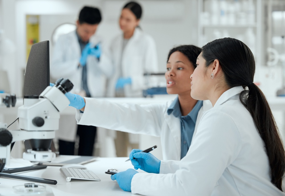
Michigan State University researchers are launching a novel project to transform how medications are developed and prescribed for women.
A US$4.6m award will fund computer models designed to predict how hormonal changes affect the way medicines move through and act in women’s bodies.
The funding is the first instalment of an award worth up to US$12.8m over three years, supporting work intended to account for hormonal changes throughout women’s lives.
Researchers plan to examine factors including menstrual cycles, pregnancy, contraceptive use, menopause and hormone replacement therapy, which can affect responses to medicines.
Teresa K. Woodruff, lead investigator on the project, said: “Because female hormone levels are constantly shifting, precision medicine allows us to map out these complex interactions.
“This NIH-backed initiative will create the first computationally driven clinical tool designed to guide medical care across every stage of a woman’s life.”
The project is led by researchers at Michigan State University and funded by the National Institutes of Health, with collaborators from Rutgers, Emory, Tulane, the University of Colorado Anschutz, the University of Michigan and the University of Utah.
Thirteen researchers will develop computational models to predict how changes in female hormones influence the way medicines move through and act within the body.
The work is part of the NIH Computational Modeling of Hormone Homeostasis Initiative, which is awarding US$21m nationally to support research into sex-specific hormonal biology.
The team plans to use artificial intelligence to digitise and organise more than 40 years of hormone research data in a publicly accessible database.
Researchers will also develop a standard computer model of a 28-day menstrual cycle, alongside models of how hormones regulate organs and tissues involved in processing nutrients, including the liver, muscle and fat.
Real-world patient data will then be used to expand the models to represent groups including women going through menopause or taking birth control, as well as women with conditions such as diabetes and obesity.
The project will also use three-dimensional human tissue models and lab-grown organoids, including liver, muscle and ovarian tissue, to test and refine the computer predictions.
Researchers plan to examine medicines including metformin, insulin and GLP-1 drugs, with the aim of developing tools that could help clinicians tailor doses and avoid harmful side effects.
Qiang Zhang, associate professor at Emory University, said: “Empowered by AI, novel assays and legacy human data, we will develop mechanistically based computational models of female physiology that can make translational, quantitative predictions for women’s responses to metabolic therapies.”
The researchers said the work could help address differences in how women respond to treatments for metabolic conditions including obesity, type 2 diabetes, cholesterol imbalances and thyroid disorders.
Nanette Santoro, professor at the University of Colorado Anschutz and president of the Endocrine Society, said: “Women experience large shifts in reproductive hormones at several points in their lifespan: puberty, pregnancy and menopause.
“During reproductive years, women also undergo profound day-to-day changes in reproductive hormone levels, giving them a markedly different endocrine backdrop than men.
“Using state-of-the-art computational technology to examine how these changes interact with commonly used medications is a critical pathway toward supporting life-course women’s health.”
The project team said its computer models and data will be made freely available to researchers and healthcare professionals when the work is completed.






 Entrepreneur2 weeks ago
KOVE Medical raises €1.7 million to improve safety of foetal surgery
 Menopause2 days ago
Menopause may not explain rising heart condition in women – study

 Fertility6 days ago
Paracetamol use may impact future fertility, studies suggest

 Sponsored Content1 week ago
Last chance to save on Women’s Health Week and Women’s Sport Summit: Early Bird pricing ends 11 September

 Menopause2 weeks ago
Menopause hormone treatment may ease brain fog, study suggests

 Menopause1 week ago
Cancer drug could tackle osteoporosis menopause weight gain
 Insight2 days ago
Women with birth trauma face 2.5x higher healthcare costs – study

 Menopause2 weeks ago
Menopausal women posing as men to buy testosterone amid NHS access issues
3 Comments