




Pregnancy
More than 80% of women iron deficient by third trimester of pregnancy


A new study and accompanying editorial has argued for routine iron screening for all pregnant women after research results revealed that more than 80 per cent of women are iron deficient by the third trimester of pregnancy.
When a woman becomes pregnant, her iron requirements increase almost tenfold to support foetal development as well as her own increased iron needs. Her ability to meet these increased iron needs depends on her iron stores at the beginning of the pregnancy as well as the physiological adaptations that enhance iron absorption as pregnancy progresses.
These physiological adaptations, however, are not always enough to support a pregnant woman’s iron needs, especially among the estimated 50 per cent of women who begin pregnancy with depleted iron stores. While often thought of as a problem in low-resource settings, recent studies have documented iron deficiency rates of 33 to 42 per cent among pregnant women in high-resource settings.
Iron deficiency can lead to anaemia, a condition in which the body can’t produce sufficient haemoglobin, which, in turn, limits the red blood cells’ ability to carry oxygenated blood throughout the body.
Anaemia during pregnancy is associated with a higher risk of both adverse maternal outcomes and adverse infant outcomes, including postpartum depression, postpartum haemorrhage, preterm birth, low birth weight, and small-for-gestational age birth. Even without the presence of anaemia, maternal iron deficiency can result in long-term neurodevelopmental challenges for the child.
At the moment, screening for iron deficiency during pregnancy is not universally routine. Moreover, there is no generally agreed upon diagnostic criteria for iron deficiency during pregnancy.
The most recent draft recommendation from the US Preventive Services Task Force, for example, states that “the current evidence is insufficient to assess the balance of benefits and harms of screening for iron deficiency anaemia in pregnant women.”
In contrast, the International Federation of Gynecology and Obstetrics and European Hematology Society recommend all pregnant women in their first trimester irrespective of the presence or absence of anaemia be screened for iron deficiency.
Moreover, they also recommend that all women of reproductive age irrespective of the presence or absence of anaemia be screened for iron deficiency.
Even when screening is conducted, it may be insufficient to detect iron deficiency. In clinical practice, for example, haemoglobin is frequently the only benchmark used to evaluate iron status among pregnant women. Haemoglobin, however only provides an indication of anaemia. As a result, poor maternal and infant health outcomes that may develop before iron deficiency advances to anaemia may arise undetected.
Unfortunately, well-designed studies of the changes in iron status during the course of pregnancy are limited.
In response, the authors of a new study evaluated the changes in iron biomarkers throughout pregnancy, established the prevalence of iron deficiency, and proposed iron status benchmarks in early pregnancy that predict iron deficiency in the third trimester.
The authors, Elaine K. McCarthy et al., also sought to determine how common risk factors for iron deficiency such as obesity and smoking affected iron status throughout pregnancy. The study is one of the largest studies ever to document the changes in iron status during pregnancy.
To conduct their research, the authors worked with data collected from 641 women in Ireland who were pregnant and had a successful delivery for the first time and who participated in the IMproved PRegnancy Outcomes via Early Detection (IMPROvED) consortium project.
Samples were taken from the women at 15 weeks, 20 weeks and 33 weeks of pregnancy to determine iron status. Within 72 hours following delivery, information about the pregnancy, delivery, and the baby were obtained from the mother via an interview with a research midwife.
Information pertaining to clinical outcomes and complications during pregnancy and delivery were confirmed by reviewing medical records.
“In this high-resource setting,” the authors found that “iron deficiency defined by a variety of biomarkers and thresholds, was very common during pregnancy, despite the cohort profile as generally healthy.”
Interestingly, none of the study participants were anaemic in the first trimester, yet more than 80 per cent of the women were iron deficient by the third trimester.
In particular, the authors noted that “our cohort had higher rates of deficiency in the third trimester than even some low-resource settings.”
In this study, almost three-quarters of the participants took an iron-containing supplement that contained the Irish/European recommended daily iron allowance of 15 to 17mg. The authors did note that “iron-containing supplements (mainly multivitamins) taken pre/early pregnancy were associated with a reduced risk of deficiency throughout pregnancy, including the third trimester.”
According to the authors, these findings draw attention to “the benefit of screening for iron deficiency with haemoglobin and ferritin in defined low-risk populations.”
Moreover, based on their findings, the authors proposed a threshold for ferritin, a protein that stores iron, of 60µg per litre or less at 15 weeks of pregnancy that predicted the presence of iron deficiency at 33 weeks of pregnancy, defined as 15µg of ferritin per litre or less.
The authors noted that: “This has previously been identified as the inflection point at which foetal iron accretion is compromised, leading to poorer neurocognitive function and earlier onset of postnatal iron deficiency in the offspring.”
In an accompanying editorial to this study, authors Michael Auerbach and Helain Landy bluntly labelled the medical community’s approach to women, including the lack of screening and treating iron deficiency and anaemia among pregnant women, as “misogyny.”
Given the study’s findings, the editorial calls upon the American College of Obstetricians and Gynecologists and the United States Preventive Services Taskforce to “change their approach to diagnosis to screen all pregnant women for iron deficiency, irrespective of the presence or absence of anaemia, and recommend supplementation when present for the most frequent nutrient deficiency disorder that we encounter.”
Looking to the future, the authors believe that “further good-quality, large-scale longitudinal studies of iron status, with concurrent inflammatory status, are needed to provide the evidence base to help establish much-needed consensus. Moreover, the use of early pregnancy iron biomarkers and thresholds should be instituted in better alignment with clinically meaningful health outcomes.”
The study and editorial have been published in The American Journal of Clinical Nutrition.



Women entering pregnancy with multiple long-term health conditions face higher risks of miscarriage and other complications, a UK study found.
Those with two or more pre-existing physical or mental health conditions had a 20 per cent higher risk of miscarriage than women with no long-term conditions.
They also had more than twice the risk of venous thromboembolism and around four times the risk of antenatal anxiety and depression.

The UK-wide research team analysed 2,225,701 pregnancies and birth events recorded between 2000 and 2022 across five datasets covering England, Scotland, Wales and Northern Ireland.
Women with multiple long-term conditions had a 69 per cent higher risk of nausea and vomiting during pregnancy and a 42 per cent higher risk of pre-eclampsia.
The women also had a 32 per cent higher risk of placental abruption and a 26 per cent higher risk of gestational diabetes.
Risks rose as the number of existing conditions increased.
Among women with three or more long-term conditions, the risk of venous thromboembolism was more than three-and-a-half times that of women with no long-term conditions.
Researchers said the findings had implications for maternity services, where care pathways are largely centred on individual conditions and may not adequately meet the needs of women with multiple long-term conditions.
Dr Kelly-Ann Eastwood, joint senior author and honorary lecturer at Queen’s University Belfast and consultant obstetrician at St Michael’s Hospital, Bristol NHS Foundation Trust, said the results “help define” the urgent clinical challenges facing women entering pregnancy with multiple long-term conditions and the clinicians caring for them across the UK.
“These findings highlight the pressing need to restructure existing maternity services to improve antenatal outcomes,” she added.
The authors cautioned that the study was observational and relied on routinely collected health records, meaning some conditions and outcomes may have been under-recorded, while residual confounding could not be excluded.
The researchers plan to examine birth and child outcomes and identify which combinations of long-term conditions carry the greatest risk.


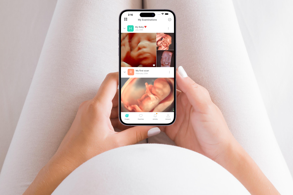
Ultrasound Direct has extended its Trice Imaging partnership for three years, adding a reporting tool across its 70-clinic UK network.
The private ultrasound provider will continue using the Tricefy platform for secure image storage and patient engagement, alongside TriceIQ for efficiency and productivity analytics.
It will also introduce Trice Workspace Reporting across its network. The companies say the tool will help standardise ultrasound reporting templates and workflows and reduce variation between clinics.
Ultrasound Direct carries out an estimated 120,000 patient scans each year across services including pregnancy, fertility, women’s health, men’s health and other diagnostic pathways.
Its network uses a large pool of sonographers working across different ultrasound systems and serves referral routes including self-referring patients, GPs and commercial partners.
Mike Steward, founding director at Ultrasound Direct, said: “Having worked with Trice Imaging since 2018, we first partnered to replace manual methods of providing scan images to expectant parents with Trice’s secure electronic image-sharing platform. Today, every study performed across the Ultrasound Direct Network is recorded and stored on Tricefy, while our clinical services have expanded considerably beyond pregnancy into fertility, women’s health, men’s health and other diagnostic pathways.”
He added: “To continue futureproofing our image management strategy across a network of 70 clinics, a large team of sonographers, varying ultrasound systems and a growing number of referral partners with different needs, we decided to extend with Trice Imaging. This includes the introduction of the new Trice Workspace Reporting module to help us scale and standardise our ultrasound reporting templates and workflow, reducing variability between clinics.”
The partnership began in 2018, initially focusing on replacing manual methods of sharing pregnancy scan images with expectant parents.
Johanna Wollert Melin, founder and chief executive of Trice Imaging Europe, said: “Ultrasound Direct has been a valued partner in the UK for 8 years.”
She added: “At the heart of the relationship is a willingness to explore new ideas, test new tools and the spirit to solve real challenges across a large and complex clinical network.”
Mark A. Samii, chief revenue officer at Trice Imaging, said: “We are delighted to retain and extend our relationship with Ultrasound Direct.”
He added: “The addition of Trice Workspace Reporting addresses a challenge we hear from multi-site providers globally – keeping reporting quality consistent across many users, systems, referral or payer relationships.”
Steward said Ultrasound Direct continues to see growing demand for private diagnostics alongside NHS care from self-referring patients and an increasingly diverse range of professional and commercial referral partners.
He added: “As that development continues, scalable digital infrastructure becomes increasingly important. Our focus is on ensuring that a growing national network can support consistent clinical workflows, different referral pathways and the technology requirements of the future, while continuing to provide patients with accessible diagnostic services.”

Paracetamol use in pregnancy was not linked to autism or ADHD, while separate research found reproductive differences in girls exposed before birth.
One study analysed health records from more than 120,000 children and found no increased risk of autism following prenatal paracetamol exposure.
A separate analysis of nearly 100,000 children also found no increased risk of ADHD among those born to mothers who used the painkiller during pregnancy.
Researchers from the Hong Kong Hospital Authority examined electronic health records covering pregnancies between January 2001 and December 2023.
The autism analysis included 124,333 children, who were nine years old on average and split almost evenly between males and females. There were 3,445 autism diagnoses, representing 2.8 per cent of the group.
The ADHD analysis involved 97,285 children, who were seven years old on average and also split evenly between males and females. There were 5,168 ADHD diagnoses, representing 5.3 per cent.
Women prescribed paracetamol during pregnancy were more likely to be older and have pre-existing conditions including psychiatric disorders, as well as reasons for taking the drug such as infection, fever or chronic pain.
No association was found between prenatal paracetamol exposure and either autism or ADHD.
The findings did not differ according to the trimester in which paracetamol was taken or whether use was intermittent or daily. Advanced maternal age, defined as pregnancy in women over 35, did not alter the findings.
The researchers wrote: “Paracetamol remains a safe and essential analgesic [pain reliever] and antipyretic [fever reducer] during pregnancy, whereas alternatives, such as NSAIDs and opioids carry well-documented risks.
“Unwarranted reluctance to use paracetamol could lead to undertreatment of pain and fever, or the use of more harmful alternatives, both posing risks to the pregnancy and developing fetus.”
The authors said women should assess paracetamol use with guidance from their doctor.
A separate study involving 685 pregnant women without pre-existing conditions and 302 infant daughters found associations between prenatal paracetamol exposure and differences in reproductive organs and hormone levels.
Researchers from Copenhagen University Hospital enrolled the women during their first trimester and assessed them during the first trimester, third trimester and again when their babies were three months old.
At around three months, infants experience a temporary rise in reproductive hormones sometimes called mini-puberty.
Pregnant participants completed questionnaires every two weeks about their use of pain medicines including paracetamol. Infant girls underwent ultrasound scans of their reproductive organs and blood tests to measure hormone levels.
Researchers also examined a separate group of 1,210 girls followed from infancy to adolescence whose mothers reported paracetamol use during the third trimester.
Three-month-old girls exposed to paracetamol before birth had an average 40 per cent smaller ovarian volume, 13 per cent smaller uterine volume and 23 per cent fewer ovarian follicles.
Girls exposed during the first trimester also had lower levels of Anti-Müllerian hormone, a marker of ovarian function.
Among the older girls, those exposed before birth were more likely to have smaller uteruses at puberty and smaller ovaries during their teenage years.
Dr Margit Bistrup Fischer, lead study author and postdoctoral researcher in the Department of Growth and Reproduction at Rigshospitalet hospital in Denmark, said: “Animal studies have demonstrated that impaired formation of ovarian follicles can lead to reduced fertility and earlier reproductive aging.
“Whether the differences observed in our study have implications for fertility and age at menopause in humans remains unknown and will require long-term follow-up of the girls in our cohort.”
She cautioned that women who had used paracetamol during pregnancy “should not be alarmed by our findings”, as the study found associations rather than direct causation and outcomes for individual women and children are unclear.
“Importantly, our study does not evaluate whether [acetaminophen] causes reproductive problems, nor does it provide evidence that prenatal exposure affects future fertility or age at menopause,” she said.
“Although we observed similar associations in an independent cohort, long-term follow-up is needed to determine whether these early-life differences have any clinical significance later in life.”









 Menopause1 week ago
Menopause may not explain rising heart condition in women – study

 Events2 weeks ago
Last chance to save on Women’s Health Week and Women’s Sport Summit: Early Bird pricing ends 11 September

 Fertility2 weeks ago
Paracetamol use may impact future fertility, studies suggest

 Mental health1 week ago
Women with birth trauma face 2.5x higher healthcare costs – study

 Menopause2 weeks ago
Cancer drug could tackle osteoporosis menopause weight gain

 News6 days ago
Medical cannabis may improve endometriosis symptoms – study

 Cancer1 week ago
Study could explain why obesity is a breast cancer risk factor

 Diagnosis2 weeks ago
FDA approves AstraZeneca breast cancer drug
1 Comment