


Fertility
IVF in transition: 2025 realities and what device manufacturers must do now

FinDBest IVF is a global B2B platform that connects manufacturers of fertility and reproductive health devices with IVF-specialist distributors in over 150 countries. We simplify global expansion, regulatory pathway planning, and distributor onboarding.
Each year, the European Society of Human Reproduction and Embryology (ESHRE) Congress reveals not just clinical updates, but also clear signals about where the IVF market is heading.
In 2025, Circular Communications compiled a focused commercial and product roadmap briefing from the event, kindly shared recently by Dr Georg Griesinger on Linkedin.
What follows is a practical breakdown of their insights—designed for medical device manufacturers and clinical users who need to make fast, evidence-based business and product decisions:
The Six Shifts Reshaping IVF
The IVF landscape in 2025 is not simply evolving—it is undergoing structural change.
Six key forces are reshaping how medical devices are adopted, evaluated, and purchased. Manufacturers who adapt early will find more predictable paths to market.
Those who do not risk falling behind as clinics tighten their criteria.
Cost pressures are now the central constraint
IVF remains financially inaccessible for large segments of the population.
In many countries, patients are still paying out of pocket.
The result is a growing preference for solutions designed around total cost of ownership (TCO).
That means not just upfront purchasing price/cost, but reusability, reliability, throughput, maintenance needs, and training time.
Products that align with capital expenditure (CAPEX) models and flexible subscriptions—especially those matched to clinic cash flow—are more likely to be adopted.
Growth in mature markets has flatlined
In many high-income countries, the number of IVF cycles per capita has plateaued.
For manufacturers, that means growth must now come from share gain or geographic expansion, particularly into fast-growing regions like Southeast Asia, the Middle East and North Africa (MENA), and Latin America.
But entering these markets successfully requires localising value propositions and working with distributors who understand IVF workflows and regulatory constraints.
Legal and ethical oversight is tightening
Questions about embryo selection, long-term storage, and artificial intelligence (AI) in diagnostics are under increased scrutiny.
For manufacturers, this raises the bar on traceability, audit readiness, and labeling compliance.
Products now need to include support for standard operating procedures (SOPs), as well as detailed logging and audit trails.
These are no longer differentiators—they are minimum requirements.
Patient experience has become a key decision factor
Clinics are under pressure to not only deliver outcomes but also reduce the emotional and cognitive burden on patients.
Devices that simplify communication, reduce the number of steps in a procedure, and help patients understand “what’s next” are increasingly favored.
Clear interfaces, intuitive indicators, and minimal user intervention all contribute to better adoption.
Clinic consolidation is shifting how buying decisions are made
Independent clinics are being replaced or absorbed by multi-site groups (Eg. US Fertility or IVIRMA, owned by KKR).
These groups prioritise enterprise-style purchasing: standardised protocols, centralised training, measurable return on investment (ROI), and clear service levels.
Manufacturers that can offer SOP kits, multi-site onboarding, and enterprise-level value metrics will have a distinct advantage.
Technology alone no longer drives differentiation
Automation, AI, microfluidics, smart incubation systems, and digital integration are becoming standard.
The key to winning adoption now lies in reproducibility, data quality, interoperability, and auditability—not just product specifications.
Clinics expect devices that integrate easily with their digital systems and produce consistent results across different settings.
Each of these shifts presents a challenge, but also a roadmap.
Cost, regulation, technology, and buyer behavior are all converging toward a more structured and evidence-driven IVF market.
Manufacturers who address these realities in their design, pricing, and commercial execution will be best positioned to scale.
Clinical and Technological Frontiers Highlighted at ESHRE 2025
Beyond the market dynamics, ESHRE 2025 spotlighted several areas of clinical innovation that are directly shaping device and diagnostics development.
These themes are not theoretical—they are influencing purchasing, adoption, and regulatory expectations now.
Ovarian stimulation protocols are being rethought As clinics aim for personalisation and patient comfort, the need for smarter diagnostics and more flexible drug delivery systems is growing.
Biomarkers that can predict ovarian reserve and treatment response are informing stimulation protocols, making room for devices that adapt to individual profiles.
At the same time, there is a clear trend toward mild stimulation protocols, which create demand for less-invasive monitoring tools and delivery systems that are intuitive, reliable, and easy to train on.
The ongoing refinement of protocols using gonadotropin-releasing hormone (GnRH) antagonists reinforces the need for workflow-agnostic solutions—those that can fit into varying cycles without adding complexity.
Embryo selection is moving well beyond morphology
Objective, evidence-backed methods are replacing subjective scoring.
One major area of interest is AI-supported time-lapse imaging, which offers the potential to assess embryo viability in a more standardised and reproducible way.
However, clinics are demanding validated tools—classified appropriately as software as a medical device (SaMD), with integration capabilities and clean clinical evidence.
In parallel, non-invasive preimplantation genetic testing (niPGT) is gaining momentum.
Media capable of capturing cell-free DNA (cfDNA), paired with ultra-sensitive genetic testing platforms, could redefine embryo selection workflows.
This is not a future trend—it’s a present R&D priority.
Manufacturers need to plan both the evidence and regulatory strategy from the outset.
Metabolomics and biomarker analysis of culture media are also being explored, particularly where kits can offer clear utility and fit easily into existing lab infrastructure.
Implantation remains a key bottleneck
Even with viable embryos, successful transfer remains challenging.
There is growing interest in non-invasive endometrial diagnostics that can assess uterine receptivity without disrupting workflow.
The market demands tools that are specific, reproducible, and easy to use.
Meanwhile, catheter design continues to influence both outcomes and patient experience.
Ergonomics, atraumatic placement, and consistent delivery are still core drivers of successful transfers.
While less discussed in marketing, this area remains a top priority for clinical users and therefore deserves more innovation attention.
Taken together, these frontiers point toward a product development path that favors integration over novelty, reproducibility over experimentation, and real-world usability over theoretical performance.
It is not just what your device does—it is how it fits into the day-to-day life of clinics under pressure.
Regulatory and Market Access: Build It In, Not On
Global regulatory expectations are rising, and shortcuts are closing.
Product teams can no longer afford to treat compliance as a post-development task. It must be embedded from Day 0.
For software and AI-based tools, classification is tightening across the United States, European Union, and China.
This means developers must create full validation plans early, align endpoints with regulatory expectations, and document cybersecurity and data governance practices before launch.
Post-market surveillance and post-market clinical follow-up are not optional; they need to be built into the development process.
Culture media and reagent products are under increased scrutiny from regulations like the European Union’s Medical Device Regulation (MDR) and In Vitro Diagnostic Regulation (IVDR).
Manufacturers must establish robust quality systems, ensure all labeling is complete and language-appropriate, and be ready to implement unique device identification requirements in every target market.
For connected lab devices, regulatory bodies expect more than just functionality.
They now require detailed documentation of data interoperability, security protocols, and integration capabilities.
Manufacturers should design clean application programming interfaces (APIs) and seamless connectors for hospital and laboratory data systems to make compliance easier—not harder—for clinics.
A practical checklist for manufacturers:
- Confirm software classification and plan validation early for each market.
- Create templates for traceability, labeling, audit logs, and PMS/PMCF.
- Implement cybersecurity and data protection frameworks from Day 0.
- Ensure unique device identification (UDI) compliance for each geography.
- Offer clear integration documents for lab systems (no assumptions).
Strategic Focus Areas for IVF Device and Diagnostics Manufacturers
- Balance cost and innovation
Demonstrate lower total cost of ownership through real-world data. Show how your product reduces maintenance, training time, or consumable use. - Support with evidence, not claims
Build prospective, multi-site clinical studies. Prepare audit-ready documentation: instructions for use, labeling, traceability, and surveillance templates. - Integrate digital and physical
Provide open, secure APIs. Ensure fast and simple onboarding for embryologists and nurses. Focus on reducing clicks, errors, and delays. - Refine embryo selection strategy
Align product claims with validated inputs—whether AI models, cfDNA media, or metabolomic markers. Monitor data drift and revalidate regularly. - Improve uterine receptivity and transfer tools
Support claims with performance data (e.g. time to placement, consistency). Offer quick training modules to accelerate adoption. - Embed regulatory design
Maintain a live matrix of requirements per SKU and market. Don’t delay planning for UDI, cybersecurity, PMS/PMCF. - Sell to enterprise buyers
Offer group-level SOP kits, ROI calculators, and centralised onboarding. Provide remote diagnostics and clear SLAs to reduce downtime. - Speed up market entry through smarter distribution
Use IVF-experienced distributors with proven regulatory capabilities. Shorten time to first order by removing the guesswork.
Key Takeaways
- Total cost of ownership is now the key metric—design around it.
- Patient workflows and clinic processes must be simplified.
- Reproducibility and integration matter more than specs.
- Plan evidence generation around the claims you want to make.
- Prepare for audits with full traceability and post-market tools.
- Offer group-ready commercial packages for multi-site chains.
- Match each market with a localised regulatory strategy.
- Choose distributors who understand both IVF and compliance.
FinDBest IVF: Your Partner in Global Expansion
These insights from ESHRE 2025, as compiled by Circular Communications, offer a compelling glimpse into the future of fertility treatment.
For medical device manufacturers, these trends are direct signals for where to focus R&D, innovation, and market entry efforts.
At FinDBest IVF, we specialise in helping medical device manufacturers navigate the complex global regulatory landscape.
Whether you’re developing cutting-edge AI for embryo selection, next-generation culture media, or advanced cryopreservation devices, we can help you:
- Find regulatory-savvy distributors and license holders.
- Identify partners who understand country-specific timelines and dossier formats.
- Expand globally, faster — with fewer surprises.
Credits
- Original, full report by Circular Communications
- Shared on Linkedin by Georg Griesinger


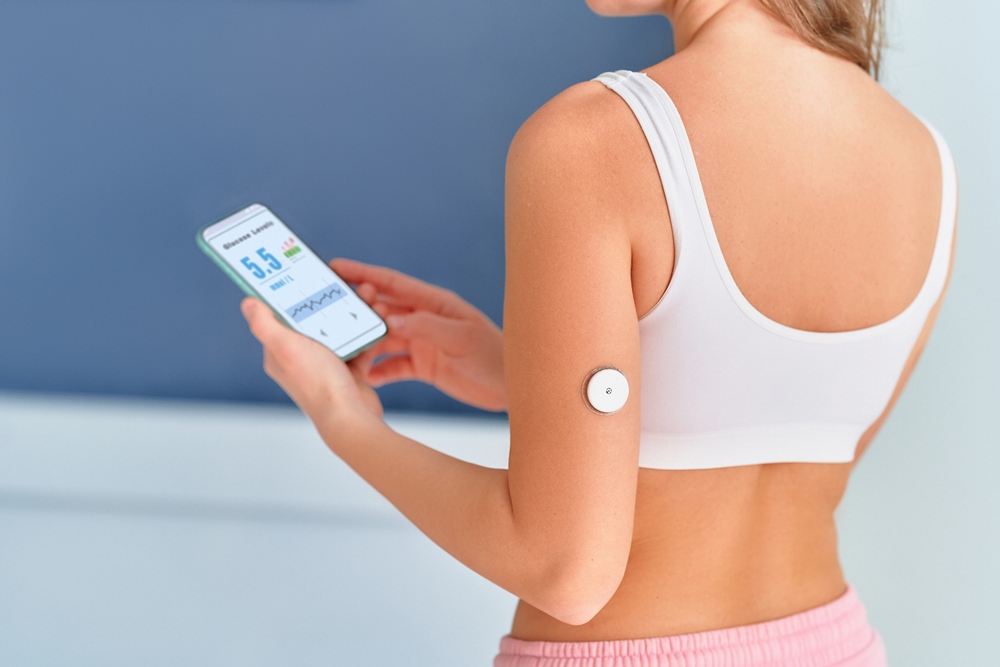
Even when standard clinical tests show normal hormone levels, men and women may have hidden problems in how their reproductive hormones are timed and coordinated, potentially affecting fertility, new research suggests.
The findings suggest reproductive health may depend not only on hormone levels in the bloodstream but also on the rhythm, timing and synchronisation of hormone changes across hours, days and the menstrual cycle.
Researchers said a wearable skin sensor patch, combined with artificial intelligence, could help detect endocrine dysfunction earlier and support more personalised fertility care.

Unexplained infertility affects about 15 to 30 per cent of couples and is diagnosed when standard investigations reveal no clear cause.
In men, current tests for infertility or hypogonadism, defined clinically as low testosterone, often include a single morning serum testosterone measurement.
In women, fertility assessment typically examines menstrual cycle characteristics and reproductive hormones such as luteinising hormone, follicle-stimulating hormone, oestradiol and progesterone.
However, reproductive hormones are not static markers. They are dynamic biological signals that rise and fall in regulated patterns throughout the day and across the menstrual cycle.
Testosterone, for example, follows a diurnal rhythm, meaning it changes across the day, while female reproductive hormones act through coordinated feedback loops involving the hypothalamic, pituitary and ovarian systems.
A single blood test may therefore miss clinically important disruption in hormonal timing.
In one study, Dr Tinatin Kutchukhidze, from the University of Oxford, examined 102 men in Georgia and the UK.
The participants were aged 22 to 38 and had normal morning total testosterone levels, measured at 12 to 35 nanomoles per litre, with or without infertility or symptoms of hypogonadism.
Hypogonadism is a condition in which the body produces too little testosterone or other sex hormones.
Kutchukhidze and colleagues used wearable AI-enabled skin sensor patches to measure testosterone levels every 15 minutes across four days.
The team found that men with symptoms had significantly disrupted testosterone rhythms, despite standard laboratory tests showing normal testosterone levels.
These previously undetected rhythm abnormalities were also associated with reduced sperm concentration and symptoms of androgen deficiency.
Androgens are hormones, including testosterone, that play an important role in reproductive health.
Kutchukhidze said: “For the first time, we have been able to track androgen patterns in real time across several days with a novel, non-invasive, continuous, AI-driven testosterone monitoring patch, compatible with Android and iPhone mobile devices.
“Previous research suggests that a normal morning testosterone level is sufficient to exclude clinically significant androgen deficiency. However, our findings challenge that assumption by demonstrating that men with normal serum testosterone may still exhibit marked disturbances in hormonal rhythmicity associated with reproductive dysfunction.”
According to the abstract, the study compared 54 men with infertility or hypogonadal symptoms with 48 age-matched healthy controls.
Mean morning serum testosterone did not differ significantly between symptomatic men and controls, at 22.4 ± 3.1 compared with 23.1 ± 3.5 nanomoles per litre.
Continuous AI-assisted monitoring, however, revealed significant differences in androgen dynamics.
Men with symptoms had lower diurnal amplitude than controls, at 5.2 ± 1.1 compared with 8.7 ± 1.4 nanomoles per litre.
The AI-derived rhythm indices predicted subclinical dysfunction with an area under the curve of 0.87, compared with 0.61 for static serum testosterone testing.
In diagnostic research, the area under the curve is used to assess how well a test distinguishes between groups, with higher values indicating stronger discrimination.
A second study by Kutchukhidze’s team examined female reproductive hormone rhythms.
The researchers developed an AI-driven metric called Endocrine Rhythm Integrity to assess whether reproductive hormones were changing in the correct pattern, at the correct time and in the correct relationship to one another across the menstrual cycle.
Endocrine refers to the hormone system, while endocrine dysfunction means hormones are not being produced or regulated in a typical way.
The team analysed data from 312 women aged 18 to 22 who had self-reported regular menstrual cycles.
Participants included fertile controls and women with unexplained infertility.
The researchers assessed key reproductive hormones during the luteal phase, including luteinising hormone, follicle-stimulating hormone, oestradiol and progesterone.
The luteal phase is the part of the menstrual cycle after ovulation. Ovulation is the release of an egg from the ovary.
They also incorporated physiological data such as basal body temperature, heart rate and sleep patterns.
Basal body temperature is the body’s resting temperature and can shift slightly around ovulation.
The study found that women with unexplained infertility had lower Endocrine Rhythm Integrity scores even when conventional hormone levels appeared normal.
Lower scores predicted infertility and were also associated with a higher incidence of implantation failure, when an embryo does not successfully attach to the womb lining.
Kutchukhidze said: “Our study reveals that a woman may have a seemingly healthy menstrual cycle and normal hormone levels but still experience hidden endocrine dysfunction that affects her ability to conceive.
“Rather than analysing hormone levels as isolated values, Endocrine Rhythm Integrity evaluates whether reproductive hormones are changing in the correct pattern, at the correct time and in the correct relationship to one another across the menstrual cycle.”
In the female study, mean cycle length did not differ significantly between fertile and infertile groups, at 28.9 ± 2.3 compared with 28.9 ± 2.5 days.
Endocrine Rhythm Integrity scores, however, were lower in the infertility group, at 0.61 ± 0.12 compared with 0.78 ± 0.10.
Disrupted endocrine rhythm integrity was observed in 64 per cent of infertile participants despite hormonally normal mid-luteal progesterone levels.
The metric independently predicted infertility status after adjustment for age, body mass index and anti-Müllerian hormone.
Anti-Müllerian hormone is made by reproductive tissues and is best known as a marker of ovarian reserve, meaning an estimate of the number of eggs remaining in the ovaries.
Receiver operating characteristic analysis indicated that Endocrine Rhythm Integrity identified infertility more effectively than cycle length or single-time-point progesterone assessment.
Lower Endocrine Rhythm Integrity scores were also associated with a higher incidence of implantation failure.
Kutchukhidze said: “Our AI-driven rhythm analyses were significantly better at identifying subclinical reproductive dysfunction than conventional testing, suggesting that both female and male endocrine disorders may not simply be disorders of hormone quantity, but rather disorders of hormonal timing, synchronisation and biological rhythm.”
The team will next assess whether the tool can reliably predict fertility outcomes across different reproductive conditions in larger and more diverse populations.


 Menopause1 week ago
Perimenopause misinformation ‘putting women at risk’

 Insight4 weeks ago
NIH Grant terminations disproportionately impact minority scientists, research finds

 Adolescent health4 weeks ago
WUKA brings Period-Positive Pool Party to London Aquatics Centre to keep girls swimming through puberty

 Insight3 weeks ago
PCOS renamed after decade-long campaign to end ‘cyst’ misconception

 Hormonal health2 weeks ago
NHS urged to update website following renaming of PCOS

 Menopause4 weeks ago
CBT shows promise for menopause insomnia and hot flashes

 Events4 weeks ago
WHIS 2026 unveils agenda and first speakers for the leading women’s health summit

 News7 days ago
Three menopause innovators shortlisted for Femtech World Award