




Cancer
MIRASOL trial shows benefits for certain ovarian cancer patients

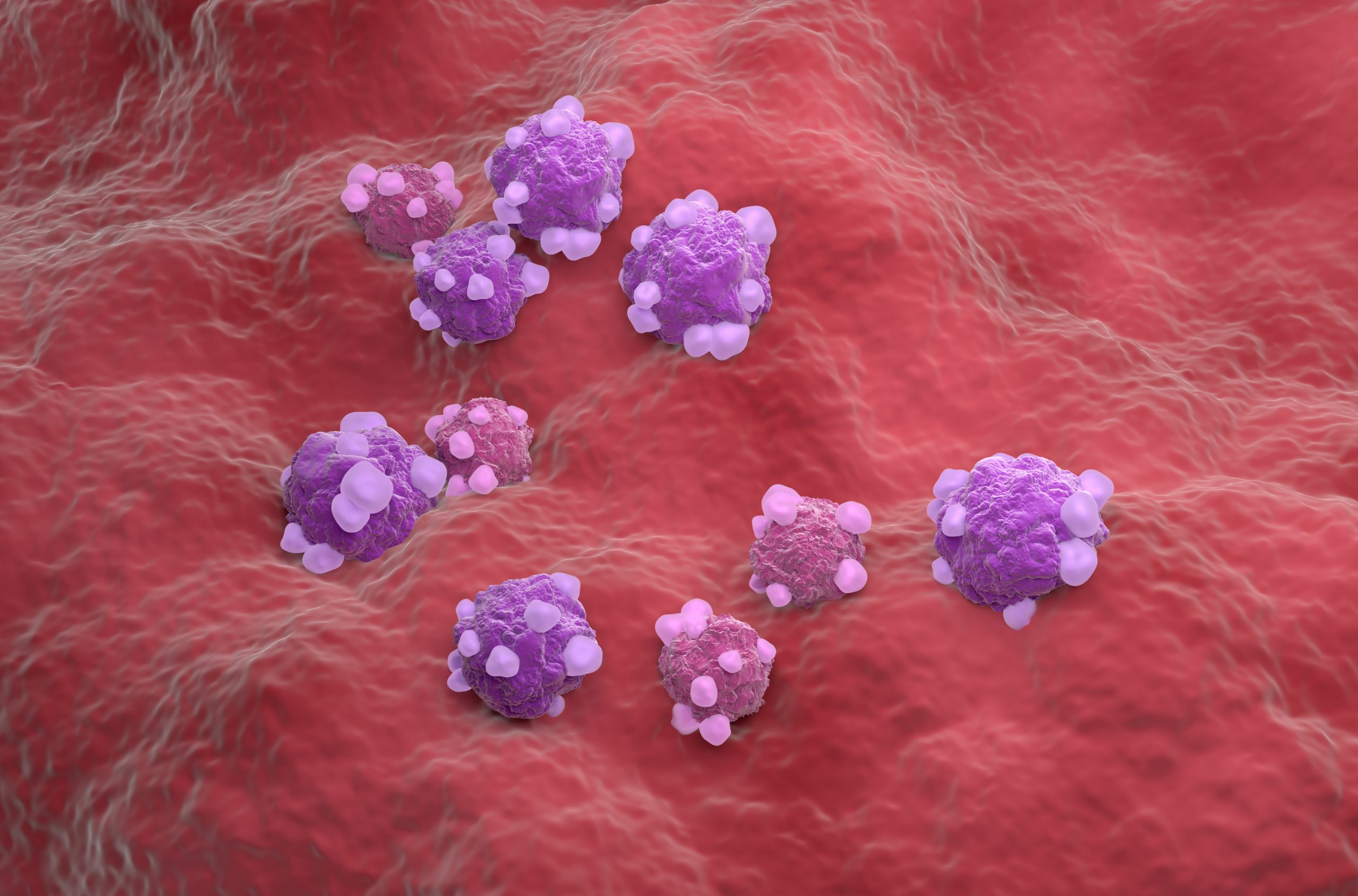
AbbVie has announced the final analysis of the confirmatory Phase 3 MIRASOL trial evaluating the efficacy and safety of ELAHERE (mirvetuximab soravtansine-gynx) in women with folate receptor alpha (FRα)-positive platinum-resistant ovarian cancer (PROC) compared to chemotherapy.
The results showed that at 30.5 months median follow-up, treatment with ELAHERE continued to show significant improvements in progression-free survival (PFS) and overall survival (OS) compared to investigator’s choice (IC) chemotherapy.
Ovarian cancer patients often present with late-stage disease and are historically first treated with platinum-based chemotherapy, which they may become resistant to and require another therapy, such as ELAHERE.

“Ovarian cancer can be devastating, and when cancer cells stop responding to chemotherapy patients may feel hopeless about their journey. The data presented today reinforce the importance of ELAHERE as a transformative therapy for patients with limited options,” said Svetlana Kobina, vice president, oncology medical affairs, AbbVie.
“We remain steadfast in our commitment to bring forward innovative therapies that improve the lives of patients with difficult-to-treat cancers.”
The Phase 3 MIRASOL study included 453 patients with high-grade serous epithelial PROC whose tumours express high levels of FRα and had been treated with up to three prior therapies.
Key findings from the 30.5-month median follow-up include that ELAHERE treatment achieved superior efficacy versus IC chemotherapy, representing a 37 per cent reduction in the risk of tumour progression or death and a higher objective response rate of 41.9 per cent versus 15.9 per cent.
It also demonstrated clinically meaningful overall survival for patients receiving ELAHERE compared to IC chemotherapy, representing a 32 per cent reduction in the risk of death.
“The final data showcase the significant improvement in overall survival benefit of treatment with ELAHERE compared to standard of care chemotherapy,” said investigator and presenter, Toon Van Gorp, professor of Gynecologic Oncology, University of Leuven.
“The significant improvements in survival, along with the well-characterized safety profile, reinforce ELAHERE as an emerging standard of care for difficult-to-treat ovarian cancer and warrants further study of this medicine in earlier treatment settings.”



A vaccine trial will test whether an mRNA jab can help stop precancerous cells developing into bowel and ovarian cancer in people with Lynch syndrome.
The first stage is due to launch this summer and will assess whether the jab can train the immune system to recognise and eliminate precancerous cells before cancer develops.
Around 175,000 people in England have Lynch syndrome, but only five per cent, or around 10,000 people, know they have it.

The inherited condition increases the risk of developing bowel cancer by 80 per cent and is linked to around 1,100 bowel cancer cases each year.
Lynch syndrome is also linked to a far higher risk of bowel, womb and ovarian cancer, alongside other types including stomach, pancreatic, kidney and skin cancer.
While the syndrome does not directly cause cancer, the genetic changes can lead to more abnormal cells developing, which then multiply and increase the risk of cancers such as bowel, prostate and endometrial cancer.
It is caused by an alteration in a mismatch repair gene. Carriers do not have any symptoms.
The new Intercept-Lynch trial is part of a scientific collaboration between the University of Oxford and Moderna, while Cancer Research UK has backed the vaccine’s development.
Once patients receive the new mRNA-4194 jab, experts will analyse their immune responses, assess the best dose and check whether the jab is safe.
The second phase of the study will include multiple centres across the UK, including Oxford, and is expected to begin in 2027.
The aim of the trial is to train the immune system with a vaccine to recognise abnormalities and stop them developing into cancer.
Professor David Church, Cancer Research UK senior cancer research fellow in the University of Oxford’s centre for human genetics and lead investigator of the trial, said: “People with Lynch syndrome are at risk of cancers over their entire lives.
“So, it’s very common, for instance, a woman to have a first cancer of her womb, and then some years later have a bowel cancer, or vice versa.
“The targets we’ve chosen for the vaccine were chosen based on their sharedness across multiple cancer types in Lynch syndrome, so we think they should provide broad protection, if the vaccine works.”
In people with Lynch syndrome, mutations can build up, making the cells containing them more likely to turn into cancerous cells.
However, those mutations can be made visible to the immune system and, with enough stimulation, the immune system can attack the abnormal cells and stop cancer from forming.
Professor Church said the mRNA jab acts as “an instruction manual” for the body to attack precancerous cells.
He added that, as with many vaccines, patients may need a booster jab at some stage.
On whether similar approaches could help prevent cancers not caused by Lynch syndrome, Professor Church said: “In terms of proof of principle that we can train the immune system to recognise these cancer-associated alterations and enhance the immune response against them to prevent these pre-cancers or prevent the progression of pre-cancer to cancer, that proof of principle should give us insights that are generalisable.”
David Berman, chief development officer at Moderna, said: “By applying mRNA technology earlier in the patient journey, we aim to harness the immune system when it can have the greatest impact.
“We are proud to bring this innovation to the UK, building on our long-standing collaboration with leading UK institutions to advance mRNA research and development.”
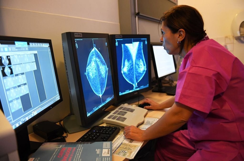
Changes in lymph nodes may help show which breast cancer patients face higher or lower risk of the disease spreading, researchers have found.
The findings could support more tailored care, new treatments and help more people avoid unnecessary treatment.
Dr Simon Vincent is chief scientific officer at Breast Cancer Now, which funded the research:
He said: “These findings suggest that changes to the structure of the lymph nodes are more than just a consequence of the cancer. They can also play an active role in helping breast cancer progress.
“With one person tragically dying from breast cancer every 45 minutes in the UK, we urgently need research like this so that we can better understand who is most at risk of their cancer progressing and becoming incurable. Only then we can find ways to stop it.
“With a better understanding of how lymph nodes change as breast cancer spreads, we could find new targets for future treatments for types of breast cancer that are harder to treat.”
Lymph nodes, a key part of the immune system, help the body fight infections and cancer. In breast cancer, the lymph nodes in the armpit are often the first place the disease spreads to.
At the moment, everyone with invasive breast cancer has to undergo surgery to remove lymph nodes so doctors can check for cancer cells.
Invasive breast cancer means cancer that has spread beyond where it first developed in the breast into nearby tissue.
While this is effective, it can lead to long-term side effects such as swelling of the arm, known as lymphoedema, and may be unnecessary for some patients, particularly those with early-stage disease or those whose cancer responds well to treatment.
The study analysed 331 lymph node samples from people with different types of breast cancer and compared them with healthy lymph nodes from people free from the disease.
It found that breast cancer could change the structure of a network that supports the lymph nodes.
Crucially, some of these changes could occur before doctors were able to spot any cancer cells in the network.
Some changes were linked to a better chance of survival, while others were associated with a poorer prognosis.
Dr Amy Llewellyn and Dr Kalnisha Naidoo from King’s College London, together with professor Sophie Acton at University College London, compared the 331 samples with healthy lymph nodes in people free from the disease.
They looked at fibroblastic reticular cells, known as FRCs, a group of cells in lymph nodes that provide their structure, control fluid flow and activate different immune cells.
The study showed that the structure of this FRC network could change before the cancer had spread and differed depending on the type of breast cancer, any spread and whether someone had received chemotherapy.
Chemotherapy uses medicines to kill cancer cells or slow their growth.
The researchers said the findings could help doctors better understand who is most at risk of breast cancer spreading.
Dr Llewellyn said the first large-scale analysis of FRC in human lymph node tissue from breast cancer patients was addressing the “urgent need” for a better understanding of the area’s biology.













 Menopause2 weeks ago
Perimenopause misinformation ‘putting women at risk’

 Hormonal health3 weeks ago
NHS urged to update website following renaming of PCOS

 News2 weeks ago
Women still being failed when they reach menopause, experts say

 Insight4 weeks ago
PCOS renamed after decade-long campaign to end ‘cyst’ misconception

 Entrepreneur2 weeks ago
Women’s Health Innovation Summit opens submissions for 2026 Innovation Showcase

 Insight1 week ago
British women among angriest in Europe, health survey reveals

 News2 weeks ago
Three menopause innovators shortlisted for Femtech World Award

 Cancer4 weeks ago
AI could transform ovarian care through personalisation, study finds