


Pregnancy
Round up: New insulin delivery technology supports healthier pregnancies and more

Femtech World explores the latest research and technology developments in the world of women’s health.
New insulin delivery technology supports healthier pregnancies
An international study has found new insulin delivery technology helps control glucose levels during pregnancy for those with Type 1 diabetes, which is crucial to the health of women and their newborns.
The technology, known as automated insulin delivery (AID), mimics a healthy pancreas. The system automatically adjusts the amount of insulin given by a pump in real-time, based on current and predicted glucose levels.
In a multicenter clinical trial, the researchers evaluated the impact of a hybrid closed-loop (HCL) insulin therapy treatment regime with standard insulin injections or an insulin pump that was not automated, along with continuous glucose monitoring.
“Keeping blood glucose in the optimal range for pregnancy is exceptionally challenging when someone has Type 1 diabetes, despite their best efforts and the support of dedicated health care clinics,” says Dr Denice Feig, MD, the study’s other co-principal investigator.
Risks associated with Type 1 diabetes in pregnancy can include increased chances of miscarriage, preeclampsia, which involves dangerous spike in blood pressure, and other significant health concerns.
Newborns of pregnant women with Type 1 diabetes are more likely to be born excessively large or early and have low blood glucose at birth and are at higher risk of birth defects.
“The study found this AID system worked in pregnancy.
“It resulted in a three hours per day improvement in the time spent in the desired glucose range compared to the standard delivery with insulin injections or regular insulin pumps,” says Donovan.
“This is very important because we have learned from other larger studies that every 72 minute per day increase, with glucose in the desired range during pregnancy, is associated with reduction in newborn complications.”
The AID system used in the study is known as a Tandem t:slim X2 insulin pump with Control-IQ technology.
The study found those using the AID spent more time in a healthy glucose level range and less time below and above the healthy range.
The improvement in blood sugar control was immediate and persisted throughout the pregnancy. These results were found at all 14 sites involved in the trial.
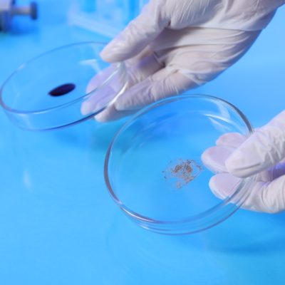
Widely-used technique for assessing IVF embryos may be flawed
A test deployed in many fertility clinics to assess the viability of embryos for use in IVF is likely to overestimate the number of embryos with abnormalities, a new study has suggested.
Using a new technique for imaging embryos in real time, a team led by scientists at the Loke Centre for Trophoblast Research, University of Cambridge, showed that abnormalities can arise at a later stage of embryo development than previously thought.
This means that the tests used in some clinics may be finding errors in cells that will go on to develop into the placenta – and abnormalities in placental cells are less likely to affect the health of the fetus.
When abnormalities are detected, the embryo may be deemed inviable and discarded, meaning patients may need to go through another cycle of treatment, which can prove costly.
So-called pre-implantation genetic testing for aneuploidy is a treatment ‘add on’ that may be offered to older women and those with a history of recurrent miscarriages or multiple IVF failures.
Researchers at the Loke Centre for Trophoblast Research, Cambridge, are interested in how early human embryos develop before implantation in the womb.
This is because in assisted conception, as many as nine in ten embryos fail to develop to a stage where they can be transferred to the womb.
To help understand development of the embryo at this early stage, Professor Niakan and colleagues, in collaboration with researchers at the Francis Crick Institute, developed a new, state-of-the-art method for watching embryos live in high resolution.
The new imaging technique involves tagging DNA inside the cell nucleus with a fluorescent protein, making it visible under a microscope.
The researchers then use an imaging technique known as light-sheet microscopy to observe the embryos in 3D as they developed without damaging them.
Of the 13 embryos analysed by the team, 10 per cent of the cells contained chromosomal abnormalities.
These arose from problems when DNA was being copied between cells, for example when chromosomes did not move properly during division or when a cell divides into three, rather than two.
Because these abnormalities arise at a relatively late stage of the embryo’s development, they appear in the outer layer of the blastocyst, which develops into the placenta – and it is from this layer that biopsies are taken for pre-genetic testing for aneuploidy.
Professor Niakan’s team is now studying cells in the inner layer to see whether such spontaneous abnormalities can also arise there.
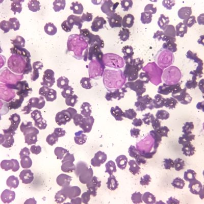
In mice, fertility treatments linked to higher mutations than natural conception
Mice pups conceived with IVF in the lab have slightly increased rates of DNA errors, or mutations, compared to pups conceived naturally.
While the results from this new study do not directly apply to humans, they highlight the importance of understanding how fertility treatments affect an offspring’s DNA.
For the study, the researchers compared genome sequences of lab mice conceived naturally and mice conceived through assisted reproductive technologies, including hormone treatments, IVF, and embryo transfer.
They discovered pups born through these fertility treatments had about 30 per cent more new single-nucleotide variants, or tiny changes in DNA sequences.
Nucleotides are DNA’s building blocks or “letters.”
Arranged in specific sequences, these letters compose the instructions cells use to grow and function.
Single-nucleotide variants are simply genetic differences (or mutations) involving a change in just one DNA letter. They can occur when cells replicate their DNA.
The mutations observed in the study are unlikely to be harmful.
Scientists estimate that fewer than 2 per cent of new mutations arising in a genome are deleterious or have an impact on an individual’s phenotype or disease susceptibility.
The mutations appeared spread across the genome, rather than clustered in particular genes.
The timing of when these new mutations appeared in early embryos also looked similar between fertility-treated and natural groups, implying that fertility treatment increases the overall chance of new DNA changes but does not impact when they occur during development.
Even with a 30 per cent increase in new mutations, the absolute number of harmful new mutations per mouse remains low.
For about every 50 mice conceived with IVF, scientists expect roughly one additional harmful DNA change compared to natural conception.
That is one problematic change out of many possible ones, since the mouse genome is about 2.7 billion DNA letters long.
A similar effect is expected if the male parent’s age increased by about 30 weeks, the researchers explained, since paternal age is a major driver of mutation rates in mammals.
The biological mechanisms underlying these genetic changes are not clear.
Further research is needed to study whether the new mutations come from a specific step in the IVF process or from the combined effects of several steps.
One possible factor is the use of hormone treatments that stimulate the ovaries, since these hormones push eggs to restart meiosis, a stage of cell division known to be prone to mistakes.
Other aspects of the fertility treatment protocol could also play a role, such as physical handling of embryos or the chemical conditions of the lab culture environment.
The study does not show whether the same effect happens in humans. Fertility procedures vary between mice and humans, and both have different reproductive biology.
For example, mice do not menstruate. Also, people seeking IVF will likely encounter environmental factors that may already have affected their genetics.


Pregnancy complications may increase women’s risk of peripheral artery disease later in life, new research suggests.
The study analysed data from more than two million women in Sweden who gave birth to single babies between 1973 and 2015.
Led by Casey Crump, the research examined the long-term risk of peripheral artery disease among women who experienced preterm delivery, pre-eclampsia, gestational diabetes or other adverse pregnancy outcomes.

Crump, professor in the department of family and community medicine at McGovern Medical School at UTHealth Houston, said: “Our prior work has already shown that adverse pregnancy outcomes are associated with long-term risks of heart disease, stroke, and heart failure.
“This study builds on that work by showing that these women have an increased risk of peripheral artery disease, an important but understudied cardiovascular condition.”
Peripheral artery disease is often a precursor to long-term cardiovascular complications, including stroke, ischaemic heart disease and premature death.
The condition affects millions of people worldwide and occurs when narrowed arteries reduce blood flow, most commonly to the legs and feet.
Ischaemic heart disease occurs when the heart does not receive enough blood and oxygen, usually because the arteries have narrowed.
Symptoms of peripheral artery disease can include leg pain, cramps while walking, numbness, cold feet and sores on the feet or legs that are slow to heal.
Crump said women who experience pregnancy complications have an important opportunity after giving birth to make plans with their primary care doctor to monitor long-term risks.
Women who have experienced complicated pregnancies should speak with their doctor about possible future cardiovascular health risks.
Checks for blood pressure, diabetes and cholesterol are important.
While the period after childbirth is an important time for women to monitor their health, Crump said it is never too late to lower the risk of peripheral artery disease.
He said: “Women with a history of adverse pregnancy outcomes who seem to be doing well may still have a higher risk that can emerge later in life.”
Crump also suggested preventive steps such as avoiding smoking, maintaining a healthy weight and following a healthy lifestyle.
He stressed the importance of long-term follow-up care and conversations with healthcare providers to help protect cardiovascular health later in life.
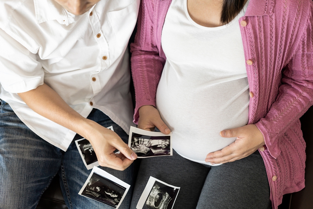
A high-powered ultrasound treatment could help identical twins affected by a rare and serious condition during early pregnancy, an initial study suggests.
Twin-to-twin transfusion syndrome, or TTTS, causes uneven blood flow between identical twins who share a placenta.
The imbalance can leave one baby dangerously small and the other too large, putting both babies’ survival at risk.

Brioney Garrett’s daughters were in danger before doctors used the world-first treatment to seal the blood vessels causing the problem without an operation.
Nancy and Margo were born healthy and, now aged four, are due to start school.
Researchers from Queen Charlotte’s and Chelsea Hospital tested the non-invasive procedure in 10 women from the UK and elsewhere in Europe after scans detected TTTS during early pregnancy.
Five women needed further treatment, while 12 of the 20 babies survived following the procedure.
The researchers described having a treatment that did not require a needle or telescope to be inserted into the mother’s abdomen as “extremely exciting”.
However, they said larger studies involving more pregnant women were needed before the procedure could be offered more widely.
Garrett described her daughters as “my miracle twins”.
She said: “We were in a very dire situation and I don’t forget that.
“It stays with me always how things could have been. Every day I still count my blessings.”
TTTS affects between 10 and 15 per cent of identical twins who share a placenta, representing around 300 to 400 pregnancies in the UK each year.
The uneven blood flow causes excess fluid to build up around the larger recipient baby, while leaving dangerously little fluid around the smaller donor baby.
Treatment usually involves inserting a needle into the womb to drain some of the fluid or using a laser to seal the connecting blood vessels.
Garrett’s procedure took about 20 minutes. She lay flat while a specially designed machine directed high-powered ultrasound waves at small blood vessels in her placenta.
She said: “It was very quick and pretty painless.”
Christoph Lees, head of fetal medicine at Imperial College Healthcare NHS Trust and professor of obstetrics at Imperial College London, described the research as “very promising”.
He said: “If this could work in a fully-fledged study, it could give hope to a lot of women who otherwise might have to have quite invasive treatment.”
Ultrasound is commonly used during medical scans to produce images of the body, but this procedure uses much more focused waves.
Heat generated by the waves can seal blood vessels about 2mm in diameter and located around 5cm to 6cm beneath the skin.
The procedure blocked blood flow in 90 per cent of the vessels treated during the study, with no unwanted side-effects reported.
Twins Trust, which supported the study, said the approach could make a significant difference for families affected by TTTS.
Helen Peck, head of healthcare engagement and research, said: “Any procedure that is non-invasive and can potentially identify TTTS earlier and improve outcomes for our families with this life-threatening condition could be a turning point.”
Scans carried out weeks after Garrett’s procedure showed that blood flow between the babies had been rebalanced, although other problems developed during the pregnancy.
Garrett said Margo, who had too little fluid around her, “was in a much better position” and that “the strain on Nancy’s heart had eased”.
Nancy and Margo were born at nearly 34 weeks, weighing 3lb 7oz and 3lb 3oz respectively.
Garrett said: “They were both healthy, and Margo wasn’t as small as we worried she was going to be.”
The twins are due to start primary school in September.
Garrett said: “They’re funny, smart, energetic little girls that just fit right in with their age group.”

A new reporting tool built specifically for obstetrics and maternal-fetal medicine has launched, aimed at teams managing increasingly complex pregnancies with limited time and resources.
Trice Imaging has released Trice Workspace Reporting, which connects imaging, reporting and longitudinal patient data in a single workflow to support faster clinical decision making.
Birth rates are falling worldwide, but pregnancies are getting more complicated. Advanced maternal age, IVF-assisted pregnancies, rising obesity rates and a higher prevalence of hypertension and diabetes mean more cases now require specialist monitoring, advanced imaging and multidisciplinary care.
At the same time, clinical teams are stretched and facing growing administrative demands.
Trice Workspace Reporting brings together customisable reporting, dynamic pregnancy dating and longitudinal patient history with an AI-ready, EHR-interoperable infrastructure, all inside the company’s Tricefy image management platform.
The company says it aims to accelerate standardised and synchronised report turnaround, support timely clinical decisions and improve operational efficiency for fetal medicine services.
“Maternal fetal medicine teams are managing increasingly complex pregnancies while being asked to do more with limited time and resources,” said Mark A. Samii, chief revenue officer at Trice Imaging.
“Trice Workspace Reporting is designed to remove unnecessary friction from reporting by creating a structured digital foundation that supports today’s need for connected clinical workflows.
“It also provides a digital foundation as practices prepare for tomorrow and the evolution of AI-enabled fetal assessment, anomaly detection and outcome prediction technologies.”
Trice Imaging describes its mission as transforming the women’s health journey by connecting physicians, patients and healthcare systems. From independent practices to large hospital ecosystems, it aims to reach the entire women’s health continuum, spanning IVF and reproductive health, maternal-fetal medicine and OB/GYN, and onwards to lifelong women’s health.
For more than 17 years the firm has worked on cloud-based storage, retrieval, display, organisation and exchange of ultrasound medical images and associated information across health environments.
Its wider platform now extends to dynamic clinical reporting, AI-driven workflow optimisation, data analytics and secure patient engagement.
Trice Imaging holds regulatory and data protection clearances in 40 countries. It has offices in Miami and Stockholm, alongside a growing network of global distributors.


 News1 week ago
New menopause drug approved for use by NHS in Scotland

 Entrepreneur2 weeks ago
Applications open for the third W Accelerate with Merck KGaA and M Ventures

 Hormonal health6 days ago
Stardust period tracker shares health data, study reveals

 News2 days ago
Zero Candida market set to reach over US$2 billion by 2030 – report

 News1 week ago
Women’s health draws record $1.55bn in equity as capital spreads beyond the mega-rounds

 Mental health2 weeks ago
SSRIs may lower heat intolerance in women with depression – study

 News2 weeks ago
Avni Wellness secures US$470k funding

 Wellness2 weeks ago
Alcohol and smoking linked to breast cancer and irregular heartbeat in women, study finds
10 Comments