


Pregnancy
Two-thirds of pregnant women miss healthy weight targets – study

Two-thirds of pregnancies involve weight gain outside recommended pregnancy weight ranges, linked to complications including preterm birth and neonatal intensive care admissions, research shows.
Analysis of 1.6 million women found only 32 per cent gained the recommended amount of weight during gestation, with 23 per cent below and 45 per cent above guidelines.
Gestational weight gain (GWG) refers to the combined growth of mother and baby during pregnancy.

The research reviewed 40 observational studies from five World Health Organization regions between 2009 and 2024.
Lead researchers Helene Teede and Rebecca Godstein say findings reinforce the need for international standards alongside lifestyle support and public health measures.
The researchers concluded: “Our findings inform and support the need for optimised, evidence-based WHO international GWG reference standards based on individual patient data, applicable across the full BMI range in contemporary and diverse global populations.
“This will build on and improve current recommendations and are essential to underpin multi-level support to improve the health of mothers and babies worldwide.”
Current guidelines from the Institute of Medicine are based on data from predominantly white women in high-income countries during the 1980s.
These do not reflect ethnically diverse populations or environmental factors driving global weight trends.
Around half (53 per cent) of participants had healthy pre-pregnancy BMI (body mass index, a measure of weight relative to height), with others classified as below (6 per cent), above (19 per cent) or obese (22 per cent).
Weight gain below recommendations was linked to lower risk of caesarean delivery and large babies but higher risk of preterm birth, small babies, low birth weight and respiratory distress.
Conversely, gaining above recommended ranges increased risks of caesarean delivery, hypertensive disorders (high blood pressure complications), large babies and neonatal intensive care admission, while lowering risks of preterm birth and small babies.
Similar patterns emerged when Asian BMI categories were applied in studies from that region.
In a linked editorial, Annick Bogaerts and Dominika Osicka said wide variation across BMI categories and regions challenges the “black and white logic” behind many clinical guidelines.
They argue for a more nuanced focus on weight gain patterns, underlying determinants and personalised, non-punitive counselling.
They wrote: “Without comprehensive, life course public health strategies, the obesity epidemic will continue across generations.
“Governments and (inter)national agencies must act now to support women’s health before, during and after pregnancy, ensuring that the next generation inherits the opportunity not the risk.”


Femtech World explores the latest research and technology developments in the world of women’s health.
New insulin delivery technology supports healthier pregnancies
An international study has found new insulin delivery technology helps control glucose levels during pregnancy for those with Type 1 diabetes, which is crucial to the health of women and their newborns.
The technology, known as automated insulin delivery (AID), mimics a healthy pancreas. The system automatically adjusts the amount of insulin given by a pump in real-time, based on current and predicted glucose levels.

In a multicenter clinical trial, the researchers evaluated the impact of a hybrid closed-loop (HCL) insulin therapy treatment regime with standard insulin injections or an insulin pump that was not automated, along with continuous glucose monitoring.
“Keeping blood glucose in the optimal range for pregnancy is exceptionally challenging when someone has Type 1 diabetes, despite their best efforts and the support of dedicated health care clinics,” says Dr Denice Feig, MD, the study’s other co-principal investigator.
Risks associated with Type 1 diabetes in pregnancy can include increased chances of miscarriage, preeclampsia, which involves dangerous spike in blood pressure, and other significant health concerns.
Newborns of pregnant women with Type 1 diabetes are more likely to be born excessively large or early and have low blood glucose at birth and are at higher risk of birth defects.
“The study found this AID system worked in pregnancy.
“It resulted in a three hours per day improvement in the time spent in the desired glucose range compared to the standard delivery with insulin injections or regular insulin pumps,” says Donovan.
“This is very important because we have learned from other larger studies that every 72 minute per day increase, with glucose in the desired range during pregnancy, is associated with reduction in newborn complications.”
The AID system used in the study is known as a Tandem t:slim X2 insulin pump with Control-IQ technology.
The study found those using the AID spent more time in a healthy glucose level range and less time below and above the healthy range.
The improvement in blood sugar control was immediate and persisted throughout the pregnancy. These results were found at all 14 sites involved in the trial.
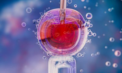
Widely-used technique for assessing IVF embryos may be flawed
A test deployed in many fertility clinics to assess the viability of embryos for use in IVF is likely to overestimate the number of embryos with abnormalities, a new study has suggested.
Using a new technique for imaging embryos in real time, a team led by scientists at the Loke Centre for Trophoblast Research, University of Cambridge, showed that abnormalities can arise at a later stage of embryo development than previously thought.
This means that the tests used in some clinics may be finding errors in cells that will go on to develop into the placenta – and abnormalities in placental cells are less likely to affect the health of the fetus.
When abnormalities are detected, the embryo may be deemed inviable and discarded, meaning patients may need to go through another cycle of treatment, which can prove costly.
So-called pre-implantation genetic testing for aneuploidy is a treatment ‘add on’ that may be offered to older women and those with a history of recurrent miscarriages or multiple IVF failures.
Researchers at the Loke Centre for Trophoblast Research, Cambridge, are interested in how early human embryos develop before implantation in the womb.
This is because in assisted conception, as many as nine in ten embryos fail to develop to a stage where they can be transferred to the womb.
To help understand development of the embryo at this early stage, Professor Niakan and colleagues, in collaboration with researchers at the Francis Crick Institute, developed a new, state-of-the-art method for watching embryos live in high resolution.
The new imaging technique involves tagging DNA inside the cell nucleus with a fluorescent protein, making it visible under a microscope.
The researchers then use an imaging technique known as light-sheet microscopy to observe the embryos in 3D as they developed without damaging them.
Of the 13 embryos analysed by the team, 10 per cent of the cells contained chromosomal abnormalities.
These arose from problems when DNA was being copied between cells, for example when chromosomes did not move properly during division or when a cell divides into three, rather than two.
Because these abnormalities arise at a relatively late stage of the embryo’s development, they appear in the outer layer of the blastocyst, which develops into the placenta – and it is from this layer that biopsies are taken for pre-genetic testing for aneuploidy.
Professor Niakan’s team is now studying cells in the inner layer to see whether such spontaneous abnormalities can also arise there.
In mice, fertility treatments linked to higher mutations than natural conception
Mice pups conceived with IVF in the lab have slightly increased rates of DNA errors, or mutations, compared to pups conceived naturally.
While the results from this new study do not directly apply to humans, they highlight the importance of understanding how fertility treatments affect an offspring’s DNA.
For the study, the researchers compared genome sequences of lab mice conceived naturally and mice conceived through assisted reproductive technologies, including hormone treatments, IVF, and embryo transfer.
They discovered pups born through these fertility treatments had about 30 per cent more new single-nucleotide variants, or tiny changes in DNA sequences.
Nucleotides are DNA’s building blocks or “letters.”
Arranged in specific sequences, these letters compose the instructions cells use to grow and function.
Single-nucleotide variants are simply genetic differences (or mutations) involving a change in just one DNA letter. They can occur when cells replicate their DNA.
The mutations observed in the study are unlikely to be harmful.
Scientists estimate that fewer than 2 per cent of new mutations arising in a genome are deleterious or have an impact on an individual’s phenotype or disease susceptibility.
The mutations appeared spread across the genome, rather than clustered in particular genes.
The timing of when these new mutations appeared in early embryos also looked similar between fertility-treated and natural groups, implying that fertility treatment increases the overall chance of new DNA changes but does not impact when they occur during development.
Even with a 30 per cent increase in new mutations, the absolute number of harmful new mutations per mouse remains low.
For about every 50 mice conceived with IVF, scientists expect roughly one additional harmful DNA change compared to natural conception.
That is one problematic change out of many possible ones, since the mouse genome is about 2.7 billion DNA letters long.
A similar effect is expected if the male parent’s age increased by about 30 weeks, the researchers explained, since paternal age is a major driver of mutation rates in mammals.
The biological mechanisms underlying these genetic changes are not clear.
Further research is needed to study whether the new mutations come from a specific step in the IVF process or from the combined effects of several steps.
One possible factor is the use of hormone treatments that stimulate the ovaries, since these hormones push eggs to restart meiosis, a stage of cell division known to be prone to mistakes.
Other aspects of the fertility treatment protocol could also play a role, such as physical handling of embryos or the chemical conditions of the lab culture environment.
The study does not show whether the same effect happens in humans. Fertility procedures vary between mice and humans, and both have different reproductive biology.
For example, mice do not menstruate. Also, people seeking IVF will likely encounter environmental factors that may already have affected their genetics.


-
 News4 weeks ago
AI embryo selection tool wins European approval
-

 News2 weeks ago
Opinion: Not ‘just stress’ – How hormonal changes affect women’s brain function
-

 Menopause4 weeks ago
Testosterone patch shows promise for menopausal women
-

 Hormonal health3 weeks ago
Top 7 drug-free solutions for managing PMS and PMDD in in 2025
-

 Entrepreneur4 weeks ago
From SEO to GEO: How women’s health brands can get found in the age of AI
-

 News4 weeks ago
FDA approves new menopause drug to treat hot flashes and night sweats
-

 Insight4 weeks ago
Spain faces protests over mammogram scandal
-

 News4 weeks ago
Most midlife women with menopause symptoms don’t seek care, research finds