




Mental health
MRI breast screening trial for hard-to-spot cancers


A new MRI breast screening trial aims to find cancers missed by mammograms in women with average breast density.
The Fast MRI Dyamond study will involve around 1,000 women aged 50 to 52 at six NHS sites, including Cheltenham, Swindon and Truro. The scans, which take around three minutes, will test whether the method can improve early detection.
Led by North Bristol NHS Trust, the study will assess whether breast cancers not visible on standard mammograms can be spotted earlier using Fast MRI.

Dr Lyn Jones, consultant radiologist and study lead, said: “We know that Fast MRI can find cancers earlier than mammograms for women with dense breasts.”
Breast density refers to the proportion of glandular to fatty tissue in the breast. On a mammogram, dense tissue appears white—similar to how cancer appears—making it harder to detect tumours.
Diana Dalgliesh, who works at Southmead Hospital, joined the three-and-a-half-year trial after losing her sister Alison to breast cancer in her 40s in 2023.
“Her breast cancer was not picked up on a mammogram which she’d had only a few months before her diagnosis,” she said.
“Perhaps if there had been a different way of screening it might have been picked up, so I thought if I am able to have an MRI scan, it may detect things earlier than if I just had a mammogram alone.”
Ms Dalgliesh said she has “little doubt” her sister’s cancer could have been found sooner with an MRI.
“I know the type of cancer she had is more easily seen on an MRI than a mammogram. Whether it would have changed the outcome I don’t know,” she added.
Fast MRI is a shorter version of the breast MRI scan already used to detect aggressive cancers under one centimetre in women with dense breast tissue.
Unlike mammograms, it does not involve compressing the breast or using radiation.
The study is jointly funded by £1.36m from the Medical Research Council and the National Institute for Health and Care Research. It is the first time women with average breast density in the UK will be offered MRI alongside routine mammogram appointments.
Janice Rose, from the patient group Independent Cancer Patients’ Voice, said the technology “could have a huge benefit to women entering the screening programme.”
“Early diagnosis for breast cancer gives women the best possible outcome,” she added.
The NHS Breast Screening Programme currently offers mammograms every three years to women aged 50 to 70. Around 1,300 lives are saved annually through early breast cancer detection.











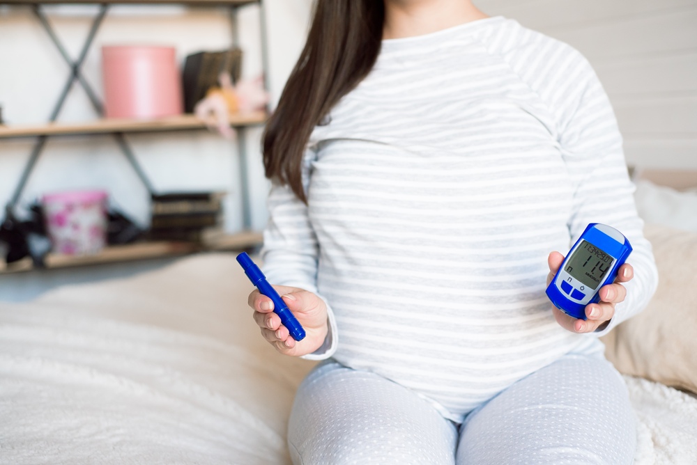
More than half of women with gestational diabetes report stigma from healthcare staff, family, friends and wider society, new research shows.
A survey of 1,800 UK women found widespread emotional distress at diagnosis of the condition, a form of high blood sugar that develops during pregnancy, with effects lasting beyond birth.
Gestational diabetes affects around one in 20 pregnancies in the UK, and the findings highlight the wider toll on women diagnosed with the condition.

The study was funded by Diabetes UK and led by researchers at King’s College London and University College Cork.
Dr Elizabeth Robertson, director of research and clinical at Diabetes UK, said: “Stigma can have a dangerous and devastating impact on pregnant women diagnosed with gestational diabetes, particularly at a time when emotions and anxieties may already be heightened.
“We know that stigma can lead to shame, isolation and poorer mental health, and may discourage people from attending healthcare appointments, potentially increasing the risk of serious complications.
“This research highlights the urgent need for better support systems, based on understanding and empathy to ensure no one feels blamed or judged during their pregnancy.”
More than two-thirds of women, 68 per cent, reported anxiety at diagnosis, while 58 per cent felt upset and 48 per cent experienced fear.
The psychological impact continued beyond birth, with 61 per cent saying the condition negatively affected their feelings about future pregnancies.
Nearly half of women, 49 per cent, felt judged for having gestational diabetes, while 47 per cent felt judged because of their body size.
More than 80 per cent felt other people did not understand gestational diabetes, and more than a third, 36 per cent, concealed their diagnosis from others.
Gestational diabetes stigma was also common in healthcare settings, with 48 per cent reporting that professionals made assumptions about their diet and exercise, and more than half, 52 per cent, feeling judged based on their blood glucose results.
Many women described a loss of control and a sense of disruption during pregnancy.
Nearly two-thirds, 64 per cent, felt they were denied a normal pregnancy, while 76 per cent reported a lack of control over their pregnancy.
More than a third, 36 per cent, felt abandoned by healthcare services after giving birth, and one in four, 25 per cent, continued to experience depression or anxiety postpartum.
Focus group participants described harmful stereotypes, including assumptions that they were ‘lazy’, had ‘poor eating habits’ or ‘lacked willpower’.
Comments from family and friends included remarks such as “should you be eating that?” and “you must have eaten too much, that’s why you have gestational diabetes.”
The researchers are calling for targeted interventions alongside structured emotional support for women during and after pregnancies affected by gestational diabetes, to improve both mental and physical health outcomes.
Professor Angus Forbes, lead researcher from King’s College London, said: “Stigma and emotional distress are far more common in women diagnosed with gestational diabetes than many realise.
“Everyday interactions, even with those who mean well, can deepen this harm, shaping women’s emotional wellbeing and the choices they feel able to make.
“It’s clear that meaningful action is needed to protect women’s mental and physical health.”
Risk factors for gestational diabetes include living with overweight or obesity, having a family history of type 2 diabetes, and being from a South Asian, Black or African Caribbean or Middle Eastern background.

Weightlifting can improve memory and mental health in older women, whether they lift heavier or lighter weights, a clinical trial has found.
The study suggests structured exercise could offer a non-drug way to help protect the ageing mind.
As people age, physical abilities often decline and the risk of cognitive impairment rises.
Women can also face a higher risk of depression and anxiety later in life because of menopause, hormonal changes and shifting social factors.
Over time, poor mental health can speed up physical and cognitive decline.
Medical professionals often recommend cardiovascular and resistance training to help preserve physical independence.
Beyond building muscle and strength, lifting weights may also help protect the brain.
The research team recruited 120 women with an average age of 68 who were not taking part in any structured exercise programmes.
Before the intervention, independent cardiologists screened the volunteers using diagnostic stress tests to make sure they could take part safely.
The researchers then divided the women into three equal groups based on their baseline physical strength to ensure a balanced comparison.
The first group followed a resistance training programme using heavier weights for eight to 12 repetitions.
The second performed the same exercises using slightly lighter weights for 10 to 15 repetitions. The third acted as a control group and remained sedentary throughout the trial.
For three months, the active groups visited the university fitness facility three mornings a week.
Under the direct supervision of qualified fitness experts, participants completed three sets of eight different full-body exercises. These included weight machines and free weights, with movements such as chest presses, leg extensions, seated rows and bicep curls.
As the women grew stronger over the 12 weeks, supervisors progressively increased the weight they lifted.
This ensured participants stayed within their assigned repetition range while maintaining proper breathing and movement technique. Researchers also told all participants not to start any new exercise outside the laboratory setting.
The scientists carried out a broad set of cognitive and psychological tests before the programme began and again shortly after it ended.
They used the Montreal Cognitive Assessment to measure spatial skills, short-term memory and language processing.
The team also used several standardised surveys to track symptoms of geriatric depression and general anxiety.
Other tests assessed executive function, the mental processes involved in planning, focusing attention and multitasking.
In the Trail Making Test, the women had to connect a scattered sequence of numbers and letters as quickly as possible to assess cognitive flexibility.
In another verbal test, they had to name as many words beginning with the letter F, or as many animals as possible, within 60 seconds.
The researchers also used a computerised Stroop test to assess inhibitory control. In this visual test, the women saw words such as “red” or “black” displayed in conflicting ink colours, such as green.
They had to suppress the automatic urge to read the word and instead press a button matching the ink colour.
After the three-month intervention, both groups of weightlifters showed clear improvements in their test scores.
Their performance on the overall cognitive assessment rose, and their reaction times in executive function tests fell substantially.
The control group showed no such improvements, and in some categories their mental performance worsened slightly.
The structured exercise also reduced the severity of mood disorders among the active participants.
Scores for depressive symptoms fell by roughly 34 per cent in the lower repetition group and 24 per cent in the higher repetition group. Anxiety scores fell by more than 40 per cent in both groups.
The researchers said these improvements met the threshold for a clinically meaningful difference.
In practical terms, that means the psychological benefits were large enough for the women to notice in their daily emotional state.
The trial found no major differences in outcomes between the two repetition strategies, suggesting both intensities worked equally well against cognitive decline.
The study has several caveats that may shape future research into the neurological benefits of structured exercise.
The testing relied heavily on self-reported psychological surveys, which can be affected by subjective bias or temporary changes in mood.
The team also did not closely track differences in the women’s light daily physical activity outside the gym.
The researchers also said the social structure of the fitness programme may have contributed to the emotional benefits.
For 12 weeks, the active participants exercised in a shared, supportive environment, with regular contact with peers and supervisors.
This kind of consistent social interaction can help reduce loneliness and provide psychological relief.
Future trials will need to isolate whether different exercise durations or extra social interaction change these positive neural effects.
Even so, the results suggest resistance training could offer an accessible way to help treat mild cognitive and mood problems.
Regular weightlifting may benefit the mind as well as the muscles in older adults.
Mental health
Poor mental health, poverty and pollution significantly raise women’s heart failure risk – study













 Entrepreneur3 weeks ago
Three sessions that show exactly where women’s health is heading in 2026

 Fertility2 days ago
Future Fertility raises Series A financing to scale AI tools redefining fertility care worldwide

 Pregnancy3 weeks ago
How NIPT has evolved and what AI NIPT means in 2026

 News3 weeks ago
Two weeks left to make your mark in women’s cardiovascular health

 Opinion4 weeks ago
Q1 momentum: Female founders are advancing, but the system still hasn’t caught up

 Mental health2 weeks ago
More research needed to understand link between brain fog and menopause, expert says

 Fertility1 week ago
Future Fertility partners with Japan’s leading IVF provider, Kato Ladies Clinic

 News2 weeks ago
Self-employment linked to better cardiovascular health outcomes in Hispanic women