


Fertility
AMH testing: the most misunderstood number in fertility – what it can and can’t tell you
Article produced in association with Spital Clinic
AMH has become one of the most-requested blood tests in private women’s health. The number it gives back is useful, but only when it is read in context.
AMH testing in the UK has gone mainstream over the past few years. Home-testing kits sell it as a snapshot of “your fertility”.
Private clinics include it in screening packages. On social media, individual AMH results are now routinely treated as a verdict on whether a woman will be able to have children.
That reading isn’t accurate. Anti-Müllerian Hormone (AMH) does carry useful information, but only inside a wider clinical picture.
Looked at on its own, it produces a lot of unnecessary anxiety, and often hides the questions that matter more.
What AMH measures
AMH is a hormone produced by the small follicles in the ovaries, the ones that haven’t yet been recruited for ovulation. Because these follicles are relatively stable across the menstrual cycle, the test can be done on any day, without needing to be timed to a period.
A higher AMH level tends to indicate a larger pool of these follicles. A lower level suggests the pool is smaller. That, broadly, is what the result shows.
The HFEA, the UK’s independent regulator of fertility treatment, describes AMH as an indicator of ovarian reserve, while making clear that fertility test results of this kind “are not guaranteed” as a predictor of fertility outcomes.
Put simply: AMH is a count of what is there. It says nothing about how well the body will use it, and it cannot predict if or when conception will happen.
Where AMH fits in a modern fertility assessment
In current UK private practice, AMH is rarely tested in isolation. A meaningful fertility assessment will pair it with a fuller hormone profile (FSH, LH, oestradiol, prolactin and thyroid function), along with markers such as Day 21 progesterone, vitamin D and rubella immunity where relevant.
This is the structure used in a trying-to-conceive screening, and there is a reason for it: each of these tests answers a different question that AMH on its own cannot.
It is this combination, not the AMH number on its own, that gives a clinician enough information to say anything meaningful about an individual’s reproductive picture.
Misconception 1: “A low AMH means natural pregnancy isn’t possible”
This is the misconception that causes the most distress, and it is consistently wrong.
Several large prospective studies of women in their 30s and 40s trying to conceive naturally have found that women whose biomarkers, including AMH, pointed to a diminished ovarian reserve were no less likely to conceive within twelve cycles than women with reassuring results.
That is why neither UK regulators nor national guidance treat AMH as a test that can predict natural fertility in women who have no known infertility issue.
The reason is simple. Natural conception only requires one good egg, released in a normal cycle, in the right window.
AMH doesn’t measure egg quality, and it doesn’t reveal whether ovulation is happening. A woman with low AMH may still ovulate every month with high-quality eggs.
A woman with high AMH (often the pattern seen in polycystic ovary syndrome) may not be ovulating regularly at all.
The NHS emphasises that age is the strongest single predictor of natural fertility. A 35-year-old with a low AMH and regular cycles is, on average, more likely to conceive naturally than a 40-year-old with a normal AMH and irregular ones.
If AMH comes back low for someone who is trying to conceive, the more useful question isn’t whether pregnancy is still possible (the answer is almost always yes), but whether there is reason to investigate the wider picture now rather than waiting twelve months.
Misconception 2: “A normal AMH means everything is fine”
The opposite assumption is just as risky.
AMH tells you about egg quantity. It does not tell you about:
- Egg quality, which is closely tied to age
- Whether ovulation is happening regularly
- Whether the fallopian tubes are open
- Whether there are structural issues such as fibroids, polyps, ovarian cysts or endometriosis
- Sperm parameters in a male partner
- Whether implantation will succeed
A reassuringly normal AMH at 38 still sits alongside age-related changes in egg quality. A slightly lower-than-average AMH at 28 may carry no real-world implications at all.
That is why no UK clinical body recommends AMH as a routine screening test for healthy women who have no fertility concerns. NICE’s fertility guideline, NG73, treats AMH as one component of a broader investigation, not as a verdict in itself.
Imaging is the natural counterpart to the blood test. A transvaginal pelvic ultrasound directly visualises the small follicles that produce AMH, the antral follicle count. It also picks up structural findings a blood test will never reveal, including ovarian cysts, fibroids, polycystic ovarian morphology, and abnormalities in the uterine cavity. A full ovarian reserve assessment normally includes both.
Where the AMH number actually matters
There are three settings in which AMH carries real, decision-relevant information.
Before IVF or egg freezing. AMH is one of the better predictors of how the ovaries are likely to respond to stimulation medication.
A higher AMH usually predicts more eggs collected per cycle, and a very low AMH may shape decisions about protocol or whether to bank cycles before treatment.
During a fertility investigation. If a couple has been trying for twelve months, or six months if the woman is over 35, AMH becomes part of a wider assessment that should also include ovarian ultrasound, a fuller hormone profile, semen analysis and an assessment of tubal patency.
As context for women planning ahead. Women who want to understand their reproductive options before they are ready to conceive (for example, ahead of a decision about egg freezing) can find AMH informative, provided it is interpreted alongside age, antral follicle count, and other markers, by a clinician who can place the number in context.
Reading the number properly
For anyone who has had an AMH test, three things make the result more useful:
- Pair it with age. A “normal” AMH at 25 means something very different from the same number at 38. Age is doing more work in the equation than the AMH value itself.
- Pair it with imaging. Ultrasound shows what is actually in the ovaries today, rather than relying on a single biochemical marker.
- Read it with a clinician. A number on a screen, with no context, no follow-up and no plan, is the worst way to use a test that, properly interpreted, can be very informative.
AMH is a useful tool. It just isn’t the headline it has often been turned into.
Disclaimer
This article is produced for informational purposes only and does not constitute medical advice, diagnosis or treatment. Clinical guidance referenced reflects published HFEA, NHS and NICE information available as at May 2026. Individual circumstances vary; readers are advised to consult a qualified healthcare professional before acting on any information in this article. This piece was produced in association with Spital Clinic, which provided background clinical information for editorial purposes. Hyperlinks to external sources are included for reference only and do not represent an endorsement of any product, service or organisation.


Even when standard clinical tests show normal hormone levels, men and women may have hidden problems in how their reproductive hormones are timed and coordinated, potentially affecting fertility, new research suggests.
The findings suggest reproductive health may depend not only on hormone levels in the bloodstream but also on the rhythm, timing and synchronisation of hormone changes across hours, days and the menstrual cycle.
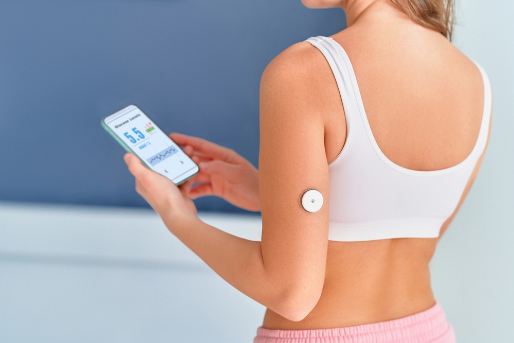
Researchers said a wearable skin sensor patch, combined with artificial intelligence, could help detect endocrine dysfunction earlier and support more personalised fertility care.

Unexplained infertility affects about 15 to 30 per cent of couples and is diagnosed when standard investigations reveal no clear cause.
In men, current tests for infertility or hypogonadism, defined clinically as low testosterone, often include a single morning serum testosterone measurement.
In women, fertility assessment typically examines menstrual cycle characteristics and reproductive hormones such as luteinising hormone, follicle-stimulating hormone, oestradiol and progesterone.
However, reproductive hormones are not static markers. They are dynamic biological signals that rise and fall in regulated patterns throughout the day and across the menstrual cycle.
Testosterone, for example, follows a diurnal rhythm, meaning it changes across the day, while female reproductive hormones act through coordinated feedback loops involving the hypothalamic, pituitary and ovarian systems.
A single blood test may therefore miss clinically important disruption in hormonal timing.
In one study, Dr Tinatin Kutchukhidze, from the University of Oxford, examined 102 men in Georgia and the UK.
The participants were aged 22 to 38 and had normal morning total testosterone levels, measured at 12 to 35 nanomoles per litre, with or without infertility or symptoms of hypogonadism.
Hypogonadism is a condition in which the body produces too little testosterone or other sex hormones.
Kutchukhidze and colleagues used wearable AI-enabled skin sensor patches to measure testosterone levels every 15 minutes across four days.
The team found that men with symptoms had significantly disrupted testosterone rhythms, despite standard laboratory tests showing normal testosterone levels.
These previously undetected rhythm abnormalities were also associated with reduced sperm concentration and symptoms of androgen deficiency.
Androgens are hormones, including testosterone, that play an important role in reproductive health.
Kutchukhidze said: “For the first time, we have been able to track androgen patterns in real time across several days with a novel, non-invasive, continuous, AI-driven testosterone monitoring patch, compatible with Android and iPhone mobile devices.
“Previous research suggests that a normal morning testosterone level is sufficient to exclude clinically significant androgen deficiency. However, our findings challenge that assumption by demonstrating that men with normal serum testosterone may still exhibit marked disturbances in hormonal rhythmicity associated with reproductive dysfunction.”
According to the abstract, the study compared 54 men with infertility or hypogonadal symptoms with 48 age-matched healthy controls.
Mean morning serum testosterone did not differ significantly between symptomatic men and controls, at 22.4 ± 3.1 compared with 23.1 ± 3.5 nanomoles per litre.
Continuous AI-assisted monitoring, however, revealed significant differences in androgen dynamics.
Men with symptoms had lower diurnal amplitude than controls, at 5.2 ± 1.1 compared with 8.7 ± 1.4 nanomoles per litre.
The AI-derived rhythm indices predicted subclinical dysfunction with an area under the curve of 0.87, compared with 0.61 for static serum testosterone testing.
In diagnostic research, the area under the curve is used to assess how well a test distinguishes between groups, with higher values indicating stronger discrimination.
A second study by Kutchukhidze’s team examined female reproductive hormone rhythms.
The researchers developed an AI-driven metric called Endocrine Rhythm Integrity to assess whether reproductive hormones were changing in the correct pattern, at the correct time and in the correct relationship to one another across the menstrual cycle.
Endocrine refers to the hormone system, while endocrine dysfunction means hormones are not being produced or regulated in a typical way.
The team analysed data from 312 women aged 18 to 22 who had self-reported regular menstrual cycles.
Participants included fertile controls and women with unexplained infertility.
The researchers assessed key reproductive hormones during the luteal phase, including luteinising hormone, follicle-stimulating hormone, oestradiol and progesterone.
The luteal phase is the part of the menstrual cycle after ovulation. Ovulation is the release of an egg from the ovary.
They also incorporated physiological data such as basal body temperature, heart rate and sleep patterns.
Basal body temperature is the body’s resting temperature and can shift slightly around ovulation.
The study found that women with unexplained infertility had lower Endocrine Rhythm Integrity scores even when conventional hormone levels appeared normal.
Lower scores predicted infertility and were also associated with a higher incidence of implantation failure, when an embryo does not successfully attach to the womb lining.
Kutchukhidze said: “Our study reveals that a woman may have a seemingly healthy menstrual cycle and normal hormone levels but still experience hidden endocrine dysfunction that affects her ability to conceive.
“Rather than analysing hormone levels as isolated values, Endocrine Rhythm Integrity evaluates whether reproductive hormones are changing in the correct pattern, at the correct time and in the correct relationship to one another across the menstrual cycle.”
In the female study, mean cycle length did not differ significantly between fertile and infertile groups, at 28.9 ± 2.3 compared with 28.9 ± 2.5 days.
Endocrine Rhythm Integrity scores, however, were lower in the infertility group, at 0.61 ± 0.12 compared with 0.78 ± 0.10.
Disrupted endocrine rhythm integrity was observed in 64 per cent of infertile participants despite hormonally normal mid-luteal progesterone levels.
The metric independently predicted infertility status after adjustment for age, body mass index and anti-Müllerian hormone.
Anti-Müllerian hormone is made by reproductive tissues and is best known as a marker of ovarian reserve, meaning an estimate of the number of eggs remaining in the ovaries.
Receiver operating characteristic analysis indicated that Endocrine Rhythm Integrity identified infertility more effectively than cycle length or single-time-point progesterone assessment.
Lower Endocrine Rhythm Integrity scores were also associated with a higher incidence of implantation failure.
Kutchukhidze said: “Our AI-driven rhythm analyses were significantly better at identifying subclinical reproductive dysfunction than conventional testing, suggesting that both female and male endocrine disorders may not simply be disorders of hormone quantity, but rather disorders of hormonal timing, synchronisation and biological rhythm.”
The team will next assess whether the tool can reliably predict fertility outcomes across different reproductive conditions in larger and more diverse populations.


 Menopause1 week ago
Perimenopause misinformation ‘putting women at risk’

 News4 weeks ago
NIH Grant terminations disproportionately impact minority scientists, research finds

 Hormonal health3 weeks ago
PCOS renamed after decade-long campaign to end ‘cyst’ misconception

 Hormonal health2 weeks ago
NHS urged to update website following renaming of PCOS

 Adolescent health4 weeks ago
WUKA brings Period-Positive Pool Party to London Aquatics Centre to keep girls swimming through puberty

 Menopause4 weeks ago
CBT shows promise for menopause insomnia and hot flashes

 Entrepreneur1 week ago
Women’s Health Innovation Summit opens submissions for 2026 Innovation Showcase

 Diagnosis4 weeks ago
Artera receives FDA Clearance for breast cancer platform