


Fertility
AMH testing: the most misunderstood number in fertility – what it can and can’t tell you
Article produced in association with Spital Clinic
AMH has become one of the most-requested blood tests in private women’s health. The number it gives back is useful, but only when it is read in context.
AMH testing in the UK has gone mainstream over the past few years. Home-testing kits sell it as a snapshot of “your fertility”.
Private clinics include it in screening packages. On social media, individual AMH results are now routinely treated as a verdict on whether a woman will be able to have children.
That reading isn’t accurate. Anti-Müllerian Hormone (AMH) does carry useful information, but only inside a wider clinical picture.
Looked at on its own, it produces a lot of unnecessary anxiety, and often hides the questions that matter more.
What AMH measures
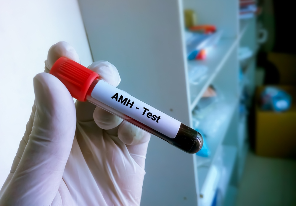
AMH is a hormone produced by the small follicles in the ovaries, the ones that haven’t yet been recruited for ovulation. Because these follicles are relatively stable across the menstrual cycle, the test can be done on any day, without needing to be timed to a period.
A higher AMH level tends to indicate a larger pool of these follicles. A lower level suggests the pool is smaller. That, broadly, is what the result shows.
The HFEA, the UK’s independent regulator of fertility treatment, describes AMH as an indicator of ovarian reserve, while making clear that fertility test results of this kind “are not guaranteed” as a predictor of fertility outcomes.
Put simply: AMH is a count of what is there. It says nothing about how well the body will use it, and it cannot predict if or when conception will happen.
Where AMH fits in a modern fertility assessment
In current UK private practice, AMH is rarely tested in isolation. A meaningful fertility assessment will pair it with a fuller hormone profile (FSH, LH, oestradiol, prolactin and thyroid function), along with markers such as Day 21 progesterone, vitamin D and rubella immunity where relevant.
This is the structure used in a trying-to-conceive screening, and there is a reason for it: each of these tests answers a different question that AMH on its own cannot.
It is this combination, not the AMH number on its own, that gives a clinician enough information to say anything meaningful about an individual’s reproductive picture.
Misconception 1: “A low AMH means natural pregnancy isn’t possible”
This is the misconception that causes the most distress, and it is consistently wrong.
Several large prospective studies of women in their 30s and 40s trying to conceive naturally have found that women whose biomarkers, including AMH, pointed to a diminished ovarian reserve were no less likely to conceive within twelve cycles than women with reassuring results.
That is why neither UK regulators nor national guidance treat AMH as a test that can predict natural fertility in women who have no known infertility issue.
The reason is simple. Natural conception only requires one good egg, released in a normal cycle, in the right window.
AMH doesn’t measure egg quality, and it doesn’t reveal whether ovulation is happening. A woman with low AMH may still ovulate every month with high-quality eggs.
A woman with high AMH (often the pattern seen in polycystic ovary syndrome) may not be ovulating regularly at all.
The NHS emphasises that age is the strongest single predictor of natural fertility. A 35-year-old with a low AMH and regular cycles is, on average, more likely to conceive naturally than a 40-year-old with a normal AMH and irregular ones.
If AMH comes back low for someone who is trying to conceive, the more useful question isn’t whether pregnancy is still possible (the answer is almost always yes), but whether there is reason to investigate the wider picture now rather than waiting twelve months.
Misconception 2: “A normal AMH means everything is fine”
The opposite assumption is just as risky.
AMH tells you about egg quantity. It does not tell you about:
- Egg quality, which is closely tied to age
- Whether ovulation is happening regularly
- Whether the fallopian tubes are open
- Whether there are structural issues such as fibroids, polyps, ovarian cysts or endometriosis
- Sperm parameters in a male partner
- Whether implantation will succeed
A reassuringly normal AMH at 38 still sits alongside age-related changes in egg quality. A slightly lower-than-average AMH at 28 may carry no real-world implications at all.
That is why no UK clinical body recommends AMH as a routine screening test for healthy women who have no fertility concerns. NICE’s fertility guideline, NG73, treats AMH as one component of a broader investigation, not as a verdict in itself.
Imaging is the natural counterpart to the blood test. A transvaginal pelvic ultrasound directly visualises the small follicles that produce AMH, the antral follicle count. It also picks up structural findings a blood test will never reveal, including ovarian cysts, fibroids, polycystic ovarian morphology, and abnormalities in the uterine cavity. A full ovarian reserve assessment normally includes both.
Where the AMH number actually matters
There are three settings in which AMH carries real, decision-relevant information.
Before IVF or egg freezing. AMH is one of the better predictors of how the ovaries are likely to respond to stimulation medication.
A higher AMH usually predicts more eggs collected per cycle, and a very low AMH may shape decisions about protocol or whether to bank cycles before treatment.
During a fertility investigation. If a couple has been trying for twelve months, or six months if the woman is over 35, AMH becomes part of a wider assessment that should also include ovarian ultrasound, a fuller hormone profile, semen analysis and an assessment of tubal patency.
As context for women planning ahead. Women who want to understand their reproductive options before they are ready to conceive (for example, ahead of a decision about egg freezing) can find AMH informative, provided it is interpreted alongside age, antral follicle count, and other markers, by a clinician who can place the number in context.
Reading the number properly
For anyone who has had an AMH test, three things make the result more useful:
- Pair it with age. A “normal” AMH at 25 means something very different from the same number at 38. Age is doing more work in the equation than the AMH value itself.
- Pair it with imaging. Ultrasound shows what is actually in the ovaries today, rather than relying on a single biochemical marker.
- Read it with a clinician. A number on a screen, with no context, no follow-up and no plan, is the worst way to use a test that, properly interpreted, can be very informative.
AMH is a useful tool. It just isn’t the headline it has often been turned into.
Disclaimer
This article is produced for informational purposes only and does not constitute medical advice, diagnosis or treatment. Clinical guidance referenced reflects published HFEA, NHS and NICE information available as at May 2026. Individual circumstances vary; readers are advised to consult a qualified healthcare professional before acting on any information in this article. This piece was produced in association with Spital Clinic, which provided background clinical information for editorial purposes. Hyperlinks to external sources are included for reference only and do not represent an endorsement of any product, service or organisation.


Onto Health has acquired Levy Health, a fertility software company providing precision diagnostics and patient intake for reproductive medicine.
The acquisition, fuelled by Onto Health’s US$20m Series A fundraise in April, supports its plan to build scalable, tech-enabled infrastructure for reproductive medicine.
Onto founder Roohi Jeelani, MD, called it the first of several moves in the company’s expansion strategy in a LinkedIn post, adding that there was “more coming soon”.

She said: “This isn’t just an acquisition, it’s proof of how we’re building Onto: physician-led, tech-enabled, and built to scale without losing the personal touch fertility patients deserve.”
Headquartered in Chicago, Onto Health combines evidence-based fertility care with artificial intelligence-driven diagnostics, clinical automation and longevity science.
AI-driven diagnostics use software to analyse patient information and support clinical decision-making, rather than replace clinicians.
Levy Health, founded in Berlin with US offices in San Francisco, helps medical providers identify endocrine disorders more quickly and helps clinics streamline fertility workups.
Endocrine disorders affect the body’s hormone system, which can influence ovulation, menstrual cycles and fertility.
Co-founder Caroline Mitterdorfer said joining Onto would expand Levy Health’s fertility care tools to more clinics and patients, helping physicians focus on patient care.
Onto opened its first clinic in Chicago in February, with plans for three more in the greater Chicago area.
The company said in April that it would use its new funding, led by Artis and Humania, to support additional operations in the US and expand into the Gulf Cooperation Council.
The Gulf Cooperation Council includes six Arab states bordering the Persian Gulf.
Softening ageing ovaries could help women remain fertile for longer, early animal research suggests.
Fertility declines with age for several reasons, including poorer egg quality, fewer ovarian follicles and the gradual stiffening of ovarian tissue.
Existing fertility treatments, including hormone therapy and in vitro fertilisation, mainly address hormonal imbalances or help eggs mature or become fertilised.

Scientists are now examining whether changing the physical structure of the ovaries could provide another route for future fertility treatments.
Stuart A. Cook, of the Cardiovascular and Metabolic Disorders Programme at Duke-National University of Singapore Medical School, published an accompanying commentary on the research.
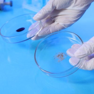
Researchers led by Shixuan Wang at Huazhong University of Science and Technology in Wuhan, China, collected healthy ovarian tissue from younger, middle-aged and older women.
They also examined samples from patients with polycystic ovary syndrome, known as PCOS, premature ovarian insufficiency, or POI, and endometriosis.
PCOS is a hormonal condition that can disrupt ovulation. POI occurs when the ovaries stop working normally before the age of 40, while endometriosis causes tissue similar to the womb lining to grow elsewhere in the body.
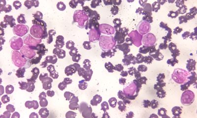
Tests of protein levels and gene activity found higher levels of the inflammatory protein interleukin-11, or IL-11, in ageing and diseased ovaries.
In laboratory experiments, the researchers exposed ovarian fibroblasts to IL-11. Fibroblasts are cells that produce connective tissue.
The protein caused the cells to produce excess collagen, a structural material that can build up during scarring and make tissue stiffer.
The researchers then genetically modified mice so they could not respond to IL-11. The animals developed less ovarian stiffening and maintained better ovarian function as they aged.
Similar results were seen in mouse models of PCOS and POI caused by chemotherapy.
In the final part of the experiment, older mice and rats were injected with a nanoparticle treatment containing small interfering RNA, or siRNA, designed to switch off IL-11.
The treatment made the animals’ ovaries less stiff and improved fertility.
Pregnancy rates among older mice rose from 25 per cent to 50 per cent, while average litter sizes also increased.
More rats treated with the therapy became pregnant and produced larger litters.
The approach remains highly speculative and will require considerably more research before its safety or effectiveness in women can be established.
However, the researchers said blocking the inflammatory pathway could eventually form the basis of new fertility treatments.
They said: “We propose that anti-IL-11 therapy represents a promising translational strategy for delaying ovarian ageing.”

W Group has opened applications for W Accelerate with Merck KGaA and M Ventures, inviting reproductive and maternal health startups, scaleups and spinouts to pitch for direct access to global pharma partnership and strategic investment.
Selected companies will pitch on 5th October, competing for the chance to accelerate their growth through commercial partnerships, investment, or both.
This is the third time Merck KGaA, a global leader in reproductive health, has partnered with W Group on the programme, which exists to close the innovation and investment gap in women’s health by connecting the sector’s most promising startups directly with the corporates and investors positioned to scale them.
What Merck KGaA and M Ventures are looking for
This year’s call is focused on breakthrough solutions in female infertility, fertility preservation, adenomyosis, endometriosis, polyendocrine metabolic ovarian syndrome (PMOS), ovarian insufficiency, preeclampsia and pregnancy comorbidities.
New for this round, applicants choose between three pathways depending on what they need from the programme:
- The Partnership Lane, for companies seeking commercial collaborations and strategic relationships
- The Investment Lane, for founders looking to connect with investors and secure funding to scale
- The Dual Lane, for innovators pursuing both partnership and investment opportunities
How the Accelerate event works
Selected companies get a 1:1 pitch practice session ahead of time, then a private 30-minute session with Merck KGaA and M Ventures leadership on the day itself, small-group sessions with regulatory and investment strategy experts, an “Ask Merck Anything” roundtable, and a VIP networking reception.
Key dates
- Open call launches: 8th July
- Open call closes: 2nd September
- Notification of successful companies: 11th September
- Pitch day: 5th October
Applications are open now at wplatform.typeform.com/to/KGzviBQM.


 Diagnosis2 weeks ago
Two “gamechanger” tests set to speed up endometriosis diagnosis on the NHS

 News7 days ago
New menopause drug approved for use by NHS in Scotland

 Cancer2 weeks ago
Thousands of women could avoid painful cancer exam with new AI blood test

 Entrepreneur2 weeks ago
Applications open for the third W Accelerate with Merck KGaA and M Ventures

 Fertility2 weeks ago
Older women face lower chance of fertility treatment working, even with donor eggs, study finds

 News2 weeks ago
We built Ema like a nurse: Here’s why that matters

 Mental health2 weeks ago
SSRIs may lower heat intolerance in women with depression – study

 Wellness2 weeks ago
Alcohol and smoking linked to breast cancer and irregular heartbeat in women, study finds