


Cancer
Tumour organoids from breast cancer patients successfully cultivated for the first time
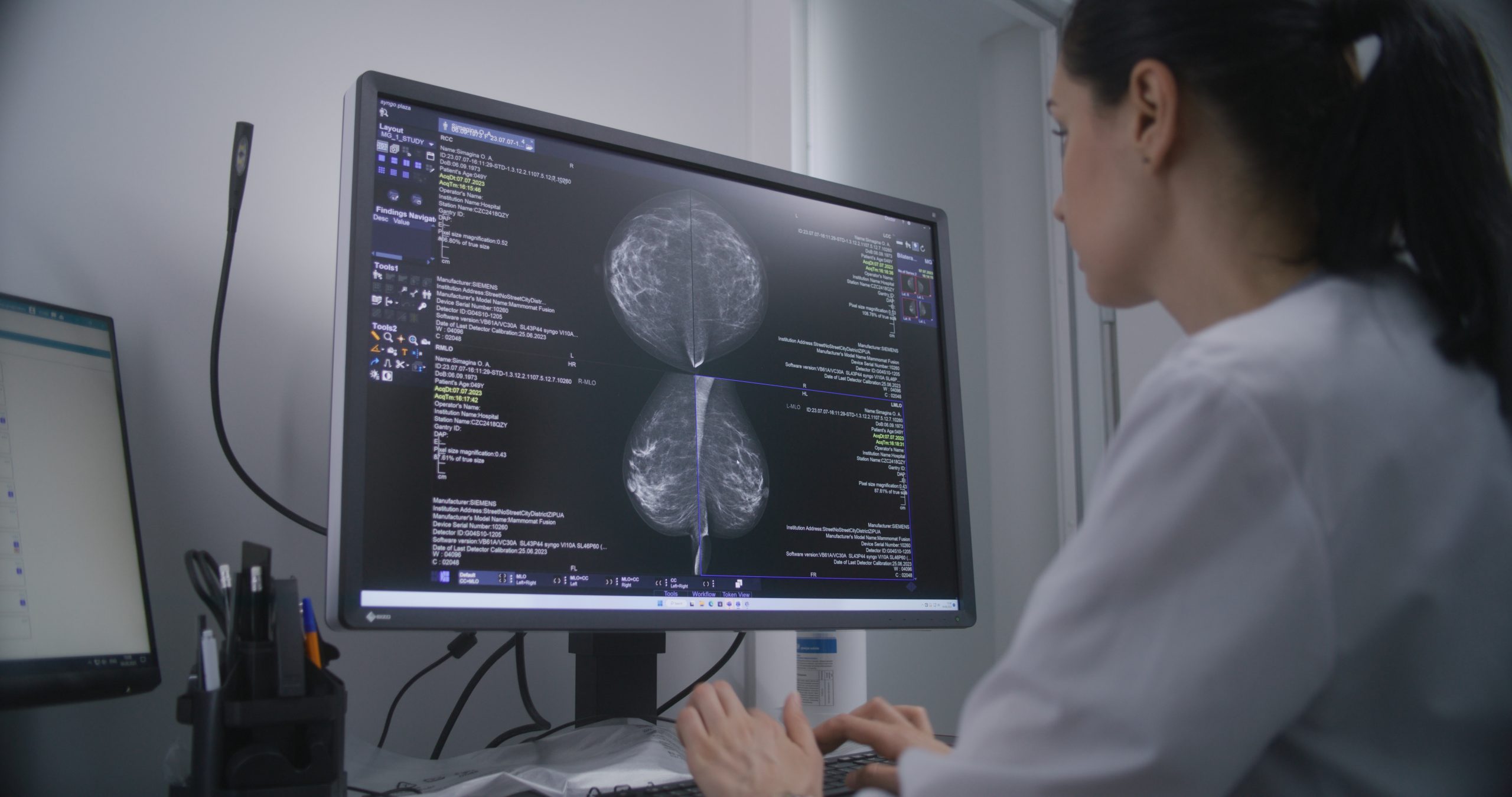
Stable tumour organoids have been directly taken from blood samples of breast cancer patients; in a world first that could speed the development of new treatments
Tumour cells circulating in the blood are the “germ cells” of breast cancer metastases – cancer cells that detach from its original tumour and spread around the body. Until now, these cells could not be propagated in culture dishes, posing a barrier to research into therapy resistance.
Using these mini-tumours, researchers have deciphered a molecular signalling pathway that ensures the cancer cells’ survival and resistance to therapy, enabling the development of an approach to eliminate them.
The researchers emphasise that these circulating cancer cells (CTCs) are extremely rare and hide among the billions of blood cells.
Andreas Trumpp, head of a research division at the German Cancer Research Center (DKFZ), which carried out the study, had previously shown that only a few CTCs can form a new metastasis in another organ.
Roberto Würth from Trumpp’s lab and first author of the paper said: “This makes it difficult to develop targeted new therapies that directly attack the metastasis-initiating cells. However, if we understand how these cells survive the initial therapy and what drives their resistance, we could tackle the formation of breast cancer metastases at the root and perhaps one day even prevent them.”
The researchers were able to multiply the CTCs and grow them as stable tumour organoids in the culture dish.
The team say that three-dimensional and patient-specific mini-tumours can be cultivated from blood samples several times during the course of the disease and are ideally suited for investigating the molecular mechanisms that enable tumours to survive despite therapy.
They found that the protein NRG1 acts like a fuel, binding to the HER3 receptor on the cancer cells and, together with the HER2 receptor, activates signalling pathways that ensure the growth and survival of the cells, and that an alternative signalling pathway, controlled by FGFR1, ensures growth and survival.
“With the help of such ‘bypasses’, tumours react to external influences, for example to targeted therapies against HER2. This is a crucial mechanism in the development of therapy resistance,” said Würth.
The researchers then used organoids to show that blocking both the NRG1-HER2/3 and FGFR signalling pathways can effectively stop the tumour growth and induce cell death.
Trumpp said: “The possibility of cultivating CTCs from the blood of breast cancer patients as tumour organoids in the laboratory at different time points is a decisive breakthrough. This makes it much easier to investigate how tumour cells become resistant to therapies.
“On this basis, we can develop new treatments that may also specifically kill resistant tumour cells. Another conceivable approach is to adapt existing therapies in such a way that the development of resistance and metastases is reduced or even prevented from the outset.
“As the organoids are specific to each patient, this method is suitable for identifying or developing customised therapies that are optimally tailored to the respective diseases.”
The team say the method must now be tested in clinical trials.


AstraZeneca’s breast cancer drug Etcamah has been approved in the EU as part of a combination treatment for advanced disease.
The European Commission acted on a positive opinion from the Committee for Medicinal Products for Human Use, the Cambridge, England-based drug maker said.

The decision followed positive results from the Serena-6 phase III trial, which showed a 56 per cent reduction in the risk of disease progression in advanced oestrogen receptor-positive breast cancer.
A phase III trial is a large, late-stage study used to assess a treatment’s safety and effectiveness before wider regulatory approval.
Oestrogen receptor-positive breast cancer is a form of the disease that can grow in response to the hormone oestrogen.
Etcamah, whose generic name is camizestrant, was tested in combination with a cyclin-dependent kinase 4/6 inhibitor.
Known as CDK4/6 inhibitors, these medicines block proteins that help cancer cells grow and divide.
AstraZeneca said the Etcamah combination has also been approved in Japan, the UAE and Saudi Arabia based on the Serena-6 trial results.
The company said breast cancer remains the leading cause of cancer death among women in Europe, with more than 140,000 deaths and more than 540,000 patients diagnosed in 2024.
AstraZeneca shares were down 0.6 per cent at 12,628 pence in London on Thursday.

Women newly diagnosed with breast cancer have a 59 per cent higher risk of ischaemic stroke in the first year after diagnosis, research suggests.
Researchers also said survivors who develop sudden stroke symptoms, including one-sided weakness, facial drooping, speech difficulties or vision loss, should seek immediate medical attention.
The multicentre study analysed National Health Insurance Service data from 107,606 women who underwent surgery for newly diagnosed breast cancer and compared them with 322,818 age-matched women with no history of cancer.

The research was conducted by professor Shin Dong-wook of Samsung Medical Center, professor Han Kyung-do of Soongsil University, professor Yong-Moon Mark Park of the University of Arkansas for Medical Sciences and professor Wonyoung Jung of the University of Pennsylvania.
Professor Yong-Moon Mark Park said: “The study demonstrates a time-dependent pattern in which the risk of ischaemic stroke rises sharply immediately after breast cancer diagnosis and treatment before gradually declining.
“The key finding is that we evaluated stroke risk according to different stages following diagnosis and treatment. This suggests that clinicians should consider not only how much the risk increases, but also when it is greatest.”
The study included women aged 18 or older who were newly diagnosed with breast cancer between 2010 and 2016, underwent surgery and had no previous stroke.
Each patient was matched with three women of the same birth year who did not have cancer. Participants were followed for an average of 7.2 years.
The main outcome was ischaemic stroke, also known as cerebral infarction. It occurs when a blocked blood vessel cuts off blood flow to the brain and is a leading cause of death and long-term disability.
During follow-up, ischaemic stroke occurred in 1,155 breast cancer patients, or 1.07 per cent, and 3,698 women in the control group, or 1.15 per cent.
Overall, breast cancer surgery was not linked to a significantly higher long-term risk of ischaemic stroke, and researchers recorded a slight fall in risk over time.
However, a different pattern emerged immediately after diagnosis.
Within one year of diagnosis, patients had a 59 per cent higher risk of ischaemic stroke than women without cancer.
The risk was highest during the first three months, at 2.90 times that of the control group.
It remained elevated within six months, at 2.27 times the control group’s risk, before gradually declining.
The risk was still 17 per cent higher three years after diagnosis.
Researchers said the temporary increase may be linked to cancer-related hypercoagulability, inflammatory responses to surgery and treatment, and cardiovascular stress caused by anticancer therapies.
Hypercoagulability means the blood is more likely than usual to form clots. Cardiovascular refers to the heart and blood vessels.
The increased risk was particularly pronounced among patients with hypertension, type 2 diabetes or a history of current smoking.
Hypertension means high blood pressure. Type 2 diabetes is a long-term condition affecting how the body controls blood sugar.
Breast cancer patients who smoked had a 2.26-fold higher risk of ischaemic stroke than comparable women without cancer.
Principal researcher professor Shin Dong-wook stressed the importance of vigilant care for patients with cardiovascular risk factors, especially during the early phase of breast cancer treatment.
Shin said: “Patients with hypertension, diabetes, or other cardiovascular risk factors, as well as those who smoke, require particularly careful management during the early phase of breast cancer treatment.
“If patients who have undergone breast cancer treatment suddenly develop weakness in one arm or leg, facial drooping, slurred or abnormal speech, or vision loss on one side, ischaemic stroke should be suspected, and they should seek immediate medical evaluation.”
Researchers said survivorship care should include strategies to monitor and manage cardiovascular and cerebrovascular disease risk throughout treatment as advances in breast cancer care continue to improve survival.
Cerebrovascular disease refers to conditions affecting blood flow and blood vessels in the brain.
Cancer cells may hijack a fertility protein to repair damaged DNA and survive chemotherapy, research suggests.
The findings could point to a way of making existing cancer treatments more effective.

SYCP1 is a protein normally involved in producing sperm and eggs.
Researchers at the University of Liverpool found that the protein, previously thought to work only in reproduction, can be reactivated in cancer cells, where it helps tumours survive and grow.
SYCP1 usually helps chromosomes pair during meiosis, the form of cell division that produces reproductive cells.
In cancer cells, however, the protein appears to take on another role. It enters the nucleus, the cell’s control centre, binds directly to DNA and regulates genes involved in cell division and DNA repair.
DNA repair is how cells fix damage to their genetic code. In cancer, this process can help tumour cells survive treatment.
The researchers found that removing SYCP1 made cancer cells much more sensitive to chemotherapy drugs that damage DNA.
The findings suggest cancers may use SYCP1 to repair damage caused by treatment and continue growing.
Dr Urszula McClurg, lecturer in biochemistry, cell and systems biology at the University of Liverpool, said: “Our findings show that cancer cells can hijack proteins that normally exist only in reproductive tissues and give them completely new jobs.
“Understanding these unexpected functions opens up exciting opportunities to develop new treatments that make existing cancer therapies more effective.”
The work challenges the long-held belief that proteins active only in fertility have no biological relevance outside the reproductive system.
Researchers say these specialised proteins could provide new treatment targets across many types of cancer.
The study also offers a new view of how cancers evolve by repurposing developmental and reproductive processes.
The findings highlight SYCP1 as a candidate for future precision cancer therapies, which are treatments based on the specific biology of a patient’s cancer.


 News2 weeks ago
New menopause drug approved for use by NHS in Scotland

 Hormonal health2 weeks ago
Stardust period tracker shares health data, study reveals

 Entrepreneur1 week ago
Zero Candida market set to reach over US$2 billion by 2030 – report

 Entrepreneur2 weeks ago
Women’s health draws record $1.55bn in equity as capital spreads beyond the mega-rounds

 Entrepreneur2 weeks ago
Onto Health acquires diagnostics software company Levy Health

 News2 weeks ago
Congress urged to invest over $20bn to close women’s health gap

 Mental health1 week ago
Nearly 60% of young women get health advice from influencers, research finds

 Insight5 days ago
From Hologic to Ark Surgical: Why Joseph LaBruzzo is betting on the future of women’s surgical innovation