


News
AHK-Cu Peptide: Implications for Dermatological Science and Beyond

The AHK-Cu peptide, a copper-binding tripeptide composed of alanine, histidine, and lysine, has sparked significant interest within various research fields, particularly in dermatological science. Emerging data suggests that this peptide, in its copper-complexed form, may exhibit remarkable properties that might revolutionize dermatological care and other research domains. The peptide’s potential extends beyond dermatology, with investigations purporting possible roles in wound healing, tissue regeneration, and anti-cellular aging. This article explores the peptide’s characteristics and its speculative implications across a range of scientific areas.
Structure and Mechanism of Action
AHK-Cu, or copper complexed with the tripeptide sequence of alanine-histidine-lysine, is an endogenously occurring biomolecule that has been found in several biological systems. Copper, a crucial trace element, is thought to play an essential role in the catalytic functions of various enzymes involved in collagen formation, antioxidant defense, and wound healing. The peptide’s primary mechanism seems to revolve around the interaction between copper and the amino acid residues, which may facilitate the copper’s exposure to target sites. These interactions might potentially lead to various cellular processes that impact tissue remodeling and regeneration.
Studies suggest that the specific tripeptide sequence that forms AHK-Cu may confer distinct molecular properties, impacting processes such as extracellular matrix synthesis, fibroblast activity, and collagen deposition. Copper ions are known for their catalytic role in many biological reactions, and the combination of AHK with copper might support the peptide’s biological functions, such as promoting the synthesis of collagen and elastin, which are paramount for maintaining the structural integrity and elasticity of the stratum corneum.
Implications in Dermatological Science
In dermatological science, AHK-Cu has attracted attention for its purported impact on deral layer regeneration and cellular anti-aging mechanisms. Research indicates that the peptide may impact the synthesis of key structural proteins like collagen, elastin, and glycosaminoglycans, all of which contribute to dermal resilience, elasticity, and hydration. The peptide’s potential to support these components may suggest its relevance in products aimed at reducing the visible signs of cellular aging, like wrinkles, sagging, and fine lines.
It has been hypothesized that the AHK-Cu peptide might encourage the proliferation and migration of skin cells, especially fibroblasts, which are critical for producing collagen and extracellular matrix components. Fibroblast stimulation may result in improved dermal texture and reduced signs of cellular aging, offering a non-invasive alternative for dermatological interventions. Moreover, research suggests that the peptide might exhibit anti-inflammatory properties, potentially providing relief from conditions associated with dermal irritation or redness.
Research indicates that AHK-Cu’s possible role in wound recovery may also hold promise in the development of cosmetic products designed to support dermal repair after injury. Investigations purport that this peptide might accelerate tissue regeneration by promoting the production of extracellular matrix components, facilitating the restoration of damaged skin structures. While still speculative, its incorporation into topical formulations might offer unique avenues for aiding post-procedure dermal layer recovery or addressing chronic dermatological conditions like eczema and psoriasis.
Regenerative Science and Wound Research
The regenerative properties of AHK-Cu are thought to extend beyond dermatological implications into the realm of wound recovery and tissue regeneration. Research indicates that the peptide may facilitate the regeneration of damaged tissues by promoting angiogenesis and the synthesis of collagen and extracellular matrix proteins. These processes are paramount for tissue repair and the formation of new blood vessels to support healing.
In research models, investigations have suggested that AHK-Cu might stimulate the migration of endothelial cells to wound sites, fostering the development of new blood vessels. This process, known as angiogenesis, is vital for supplying oxygen and nutrients to the healing tissue, thus accelerating recovery. Furthermore, investigations purport that the peptide might impact the differentiation of stem cells, potentially aiding in the regeneration of damaged tissues and organs. This may pave the way for novel approaches to wound care and tissue engineering, particularly in chronic conditions where healing is impaired.
Inflammation and Dermatological Conditions Research
Inflammation is a paramount factor in many dermal conditions, including acne, eczema, and psoriasis. The findings imply that the AHK-Cu peptide may possess anti-inflammatory properties that may be relevant in modulating the inflammatory response in these conditions. Scientists speculate that by inhibiting the production of pro-inflammatory cytokines or reducing oxidative stress, AHK-Cu might help alleviate symptoms associated with chronic dermatological conditions. Its potential to restore balance in the dermal layer’s immune response might make it an attractive candidate for future research options targeting inflammatory dermatological disorders.
Additionally, the peptide has been hypothesized to have an impact on the dermal layer’s ability to combat oxidative stress. Research indicates that copper is a critical component of antioxidant enzymes like superoxide dismutase (SOD), which protect cells from oxidative damage. It has been theorized that by stabilizing copper in its active form, AHK-Cu might contribute to better-supported antioxidant defense within the dermal layer, potentially preventing cellular damage that accelerates the cellular aging process and contributes to the development of various dermal conditions.
AHK-Cu in Hair Research
Another intriguing potential implication of AHK-Cu is its possible impact on hair growth and regeneration. Investigations purport that the peptide, in combination with copper, might stimulate the growth of hair follicles in research models by encouraging the anagen (growth) phase of the hair cycle. Copper’s role in collagen formation and its involvement in enzymatic processes linked to hair follicle health make it a critical element in hair regeneration.
Potential Relevance to Tissue Research
Beyond dermatological implications, AHK-Cu may also suggest promise in tissue engineering. This interdisciplinary field focuses on developing materials and strategies for regenerating damaged tissues and organs. The peptide’s potential to impact collagen synthesis and angiogenesis suggests its potential relevance to the creation of tissue scaffolds designed to promote cell growth and tissue regeneration.
The Future of AHK-Cu Research
The continued investigation into AHK-Cu’s properties is essential to fully understanding its potential implications. As the peptide’s mechanisms are further elucidated, new strategies may emerge for leveraging its regenerative and dermal layer-supporting properties across various scientific disciplines.
Conclusion
The AHK-Cu peptide represents a fascinating biomolecule with a range of possible implications across multiple research domains. Its potential impact on dermatologic science, wound healing, hair regeneration, and tissue engineering underscores the versatility and promise of this copper-complexed peptide. While much of the research remains speculative, the current body of knowledge points to exciting possibilities for AHK-Cu’s role in supporting dermal science, promoting tissue repair, and even regenerating hair follicles. As investigations continue, the peptide’s implications might extend even further, paving the way for innovative approaches to a variety of conditions in both specifically dermatological and other relevant scientific settings. Researchers may find the highest-quality research compounds here.
References
[i] Allen, M. D., & Wells, P. A. (2019). Copper-binding peptides and their role in skin regeneration: Insights into the mechanisms of AHK-Cu. Journal of Cosmetic Dermatology, 18(4), 1127-1135. https://doi.org/10.1111/jocd.12945
[ii] Gupta, S., & Kaur, G. (2021). AHK-Cu peptide in wound healing and tissue regeneration: Mechanisms and therapeutic applications. Regenerative Medicine, 16(3), 245-257. https://doi.org/10.1016/j.regmed.2021.03.008
[iii] Patel, M. D., & Shaw, D. J. (2020). The anti-inflammatory and antioxidant properties of AHK-Cu in dermatological disorders. Journal of Dermatological Science, 99(2), 103-111. https://doi.org/10.1016/j.jdermsci.2020.04.001
[iv] Chen, L., & Liu, F. (2022). AHK-Cu peptide and its role in hair follicle regeneration: A promising tool for hair restoration therapies. Journal of Dermatology and Hair Research, 7(1), 52-61. https://doi.org/10.1097/DHR.0000000000000294
[v] Joffe, C. R., & Lee, H. S. (2021). The potential of AHK-Cu peptide in tissue engineering: Applications in collagen synthesis and regenerative medicine. Tissue Engineering Part B: Reviews, 27(4), 379-392. https://doi.org/10.1089/ten.TEB.2021.0095

Light exercise and less sitting may reduce pregnant women’s risk of serious blood pressure complications, according to a new study.
Researchers have proposed a daily activity and sleep guide that they say was linked to a nearly 30 per cent lower risk of hypertensive disorders of pregnancy.
The suggested pattern includes fewer than eight hours of sedentary time, at least seven hours of light physical activity, around 22 minutes of more intense activity and nearly nine hours of sleep.

The University of Iowa-led study examined the daily behaviours of 470 pregnant women across all stages of pregnancy.
Participants wore monitors that measured physical activity over 24-hour periods and recorded how long they spent asleep.
Hypertensive disorders of pregnancy include chronic high blood pressure, gestational hypertension and pre-eclampsia.
Gestational hypertension is high blood pressure that develops during pregnancy, while pre-eclampsia is a potentially serious condition involving high blood pressure and signs that organs may be affected.
Sedentary behaviour means being mostly inactive, such as sitting or lying down.
Light physical activity can include casual walking, moving around the home or standing.
Moderate to vigorous activity includes movement such as brisk walking, where breathing and heart rate increase.
Kara Whitaker, associate professor in the department of health, sport, and human physiology at Iowa and corresponding author of the study, said: “We are identifying the optimal composition of movement behaviours across the day associated with the lowest risk of developing HDP and the most improved health outcomes.
“This blueprint holds for each and every trimester of pregnancy.”
Study participants were enrolled at sites in Iowa City, Pittsburgh and Morgantown, West Virginia.
The women wore activity and sleep monitors for at least one week during each trimester of pregnancy.
Four in five participants were non-Hispanic white and nearly a quarter lived in rural areas.
The data showed a steep rise in risk among pregnant women who were sedentary for more than 10 hours a day.
Women who increased light physical activity to at least four hours a day reduced their risk of hypertensive disorders of pregnancy to 15 per cent from 30 per cent.
Whitaker said: “Just moving around more seems to have significant health benefits.
“And I think it also may be a more feasible target for women who are pregnant who are not exercising regularly.”
The researchers said they were surprised that longer durations of moderate to vigorous physical activity did not appear to provide additional benefit.
Sleep beyond a certain duration also did not appear to bring major further benefits.
Whitaker said: “Through this study, we are providing evidence that reducing sedentary behaviour and engaging in light physical activity are important, and maybe more important, when it comes to pregnancy and health.”
The findings may be relevant beyond pregnancy because clinical research has shown that women who develop hypertensive disorders of pregnancy are more than twice as likely to develop heart disease later in life.
Cardiovascular disease includes conditions affecting the heart and blood vessels, such as heart disease and stroke.
Whitaker said: “We know that cardiovascular disease is the number one killer of women, and if we can intervene in pregnancy and prevent women from developing a hypertensive disorder of pregnancy, we are putting them on a better trajectory, away from cardiovascular disease and toward more optimal cardiovascular health.”
The study was published online on June 10.
A second study, published online on May 27, looked more closely at the ratio and type of sedentary behaviour and light physical activity linked to a lower risk of hypertensive disorders of pregnancy.
Whitaker is a lead co-author on that study.
Co-authors in the June 10 study include Alex Crisp, Jaemyung Kim, Karina Smith, Donna Santillan, Mark Santillan and Bridget Zimmerman, from Iowa; Jacob Gallagher, from Iowa State University; Melissa Jones, from Oakland University in Michigan; Bethany Barone Gibbs, Katrina Wilhite, Alexis Thrower and Iqra Sheikh, from West Virginia University; and Sabera Rahman, Janet Catov, Christopher Kline and Maisa Feghali, from the University of Pittsburgh.
The National Institutes of Health, the University of Iowa Institute for Clinical and Translational Science, the University of Pittsburgh Clinical and Translational Science Institute and the West Virginia Clinical and Translational Science Institute funded the research.

We are excited to reveal the winners of the third annual Femtech World Awards.
The winners were announced at a virtual event this afternoon attended by shortlisted companies, along with sponsors and judges.
The event welcomed guests from the UK, Europe, Asia, Africa and North America.
Thank you to all 174 entries, as well as the sponsors for making the event possible.
See you in 2027!
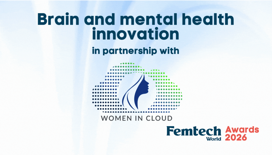
Femtech World Awards 2026 Winners
Winner:

Shortlisted:
IVI RMA x Juno Genetics
Natural Cycles

Winner:

Highly commended:
U-Ploid
Shortlisted:
Hello Inside
Winner:
![]()
WISE HF, led by Prof. Mary Ryder
Highly commended:
Cardiac College for Women
Shortlisted:
Hyvelle Ferguson-Davis
CognitiveCare

Winner:

Highly commended:
Youterus
Shortlisted:
ŌURA
Winner:

Shortlisted:
LeanShield by ParrotPal Group
Perigen
Winner:
![]()
Shortlisted:
Body Moody
Looop

Winner:
![]()
Shortlisted:
Owning Your Menopause
Womeno

Winner:

Shortlisted:
The Blue Box
Celbrea
Winner:

Shortlisted:
HealCycle
Mor

Winner:

Shortlisted:
HRC Fertility
Mira

Pregnant women’s expectations about postpartum sleep may predict sleep quality after birth, outweighing prior sleep and psychiatric history, a study suggests.
The findings suggest attitudes and beliefs about sleep during pregnancy could be a modifiable risk factor for postpartum sleep concerns.
They also indicate that, among women expecting the poorest sleep, higher postpartum anxiety may further worsen sleep quality.
Sammy Dhaliwal, lead author is clinical health psychologist and research fellow in the department of obstetrics and gynaecology at the Perelman School of Medicine at the University of Pennsylvania.
Dhaliwal said: “Most pregnant women in our sample anticipated poor postpartum sleep before it occurred, and it was striking that those expectations predicted worse sleep outcomes even after accounting for factors such as prior sleep disorders, psychiatric history, and number of previous births.
“This suggests that attitudes and beliefs about sleep during pregnancy may represent a modifiable target for early intervention before postpartum sleep problems emerge.”
Sleep disturbance affects an estimated 60 to 80 per cent of postpartum women and is linked to a higher risk of depression and anxiety.
Researchers said it is often regarded as an expected part of life after childbirth rather than a health issue that may be addressed earlier.
The study enrolled 432 pregnant women at about 24 weeks of gestation, meaning around 24 weeks into pregnancy.
Participants completed measures of their expectations about postpartum sleep, current sleep quality using the Pittsburgh Sleep Quality Index, and mood using validated depression and anxiety scales.
Assessments were repeated at six, 12 and 24 weeks postpartum.
A subset of 49 women also wore wrist actigraphy devices at six to eight weeks postpartum.
Actigraphy uses a wearable device, similar to a watch, to estimate sleep and wake patterns based on movement.
The results showed that 70 per cent of pregnant women, or 301 of 432 participants, expected poor sleep in the postpartum period.
Researchers found that predicted sleep disruption during pregnancy was a significant predictor of postpartum sleep concerns.
Among first-time pregnant women without prior health concerns, those who expected greater sleep disturbance had significantly more disrupted sleep after birth, measured by both actigraphy and self-report.
Among women who expected the worst sleep quality, higher postpartum anxiety significantly worsened both measured sleep and self-reported sleep, independent of anxiety levels during pregnancy.
Dhaliwal said the findings point to two possible areas for intervention: addressing sleep-related beliefs during pregnancy and treating postpartum anxiety.
Dhaliwal said: “Postpartum sleep disruption is often treated only after problems develop, but our findings suggest there may be an opportunity to intervene earlier during pregnancy.
“Addressing sleep-related beliefs and postpartum anxiety during prenatal and postpartum care may help improve sleep and emotional well-being in new mothers.”


 Menopause2 weeks ago
Perimenopause misinformation ‘putting women at risk’

 Hormonal health3 weeks ago
NHS urged to update website following renaming of PCOS

 News2 weeks ago
Women still being failed when they reach menopause, experts say

 Entrepreneur2 weeks ago
Women’s Health Innovation Summit opens submissions for 2026 Innovation Showcase

 Insight1 week ago
British women among angriest in Europe, health survey reveals

 News2 weeks ago
Three menopause innovators shortlisted for Femtech World Award

 Fertility4 weeks ago
AI could transform ovarian care through personalisation, study finds

 News3 weeks ago
Low insulin diet and avoiding four food groups may prevent menopause weight gain