


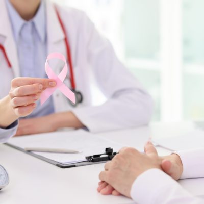
Cancer
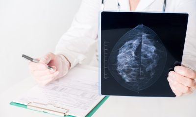
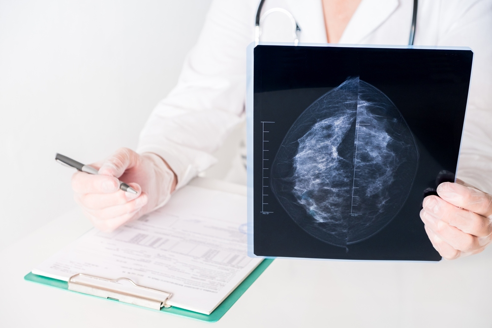
Analysing multiple mammograms improves breast cancer risk prediction

A new study describes an innovative method of analysing mammograms that significantly improves the accuracy of predicting the risk of breast cancer development over the following five years.
Using up to three years of previous mammograms, the new method identified individuals at high risk of developing breast cancer 2.3 times more accurately than the standard method, which is based on questionnaires assessing clinical risk factors alone, such as age, race and family history of breast cancer.
“We are seeking ways to improve early detection, since that increases the chances of successful treatment,” said senior author Graham Colditz, associate director of Siteman Cancer Center.
“This improved prediction of risk also may help research surrounding prevention, so that we can find better ways for women who fall into the high-risk category to lower their five-year risk of developing breast cancer.”
This risk-prediction method builds on past research led by Colditz and lead author Shu Jiang, a statistician, data scientist and associate professor of surgery in the Division of Public Health Sciences at WashU Medicine.
The researchers showed that prior mammograms hold a wealth of information on early signs of breast cancer development that can’t be perceived even by a well-trained human eye. This information includes subtle changes over time in breast density, which is a measure of the relative amounts of fibrous versus fatty tissue in the breasts.
For the new study, the team built an algorithm based on artificial intelligence that can discern subtle differences in mammograms and help identify those women at highest risk of developing a new breast tumour over a specific timeframe.
In addition to breast density, their machine-learning tool considers changes in other patterns in the images, including in texture, calcification and asymmetry within the breasts.
“Our new method is able to detect subtle changes over time in repeated mammogram images that are not visible to the eye,” said Jiang, yet these changes hold rich information that can help identify high-risk individuals.
At the moment, risk-reduction options are limited and can include drugs such as tamoxifen that lower risk but may have unwanted side effects. Most of the time, women at high risk are offered more frequent screening or the option of adding another imaging method, such as an MRI, to try to identify cancer as early as possible.
“Today, we don’t have a way to know who is likely to develop breast cancer in the future based on their mammogram images,” said co-author Debbie Bennett, associate professor of radiology and chief of breast imaging for the Mallinckrodt Institute of Radiology at WashU Medicine.
“What’s so exciting about this research is that it indicates that it is possible to glean this information from current and prior mammograms using this algorithm. The prediction is never going to be perfect, but this study suggests the new algorithm is much better than our current methods.”
AI improves prediction of breast cancer development
The researchers trained their machine-learning algorithm on the mammograms of more than 10,000 women who received breast cancer screenings through Siteman Cancer Center from 2008 – 2012. These individuals were followed through 2020, and in that time 478 were diagnosed with breast cancer.
The researchers then applied their method to predict breast cancer risk in a separate set of patients — more than 18,000 women who received mammograms through Emory University in the Atlanta area from 2013 – 2020. Subsequently, 332 women were diagnosed with breast cancer during the follow-up period, which ended in 2020.
According to the new prediction model, women in the high-risk group were 21 times more likely to be diagnosed with breast cancer over the following five years than were those in the lowest-risk group.
In the high-risk group, 53 out of every 1,000 women screened developed breast cancer over the next five years. In contrast, in the low-risk group, 2.6 women per 1,000 screened developed breast cancer over the following five years.
Under the old questionnaire-based methods, only 23 women per 1,000 screened were correctly classified in the high-risk group, providing evidence that the old method, in this case, missed 30 breast cancer cases that the new method found.
The mammograms were conducted at academic medical centres and community clinics, demonstrating that the accuracy of the method holds up in diverse settings.
Importantly, the algorithm was built with robust representation of Black women, who are usually underrepresented in development of breast cancer risk models. The accuracy for predicting risk held up across racial groups. Of the women screened through Siteman, most were white, and 27 per cent were Black. Of those screened through Emory, 42 per cent were Black.
In ongoing work, the researchers are testing the algorithm in women of diverse racial and ethnic backgrounds, including those of Asian, southeast Asian and Native American descent, to help ensure that the method is equally accurate for everyone.
The researchers are working with WashU’s Office of Technology Management toward patents and licensing on the new method with the goal of making it broadly available anywhere screening mammograms are provided. Colditz and Jiang also are working toward founding a start-up company around this technology.


The Welsh Government has been urged to invest in breast cancer services after figures showed continuing delays in diagnosis and treatment.
Breast Cancer Now said the target for 75 per cent of patients to be seen within 62 days of initial referral has been met only three times since the Suspected Cancer Pathway was introduced in 2020.
The latest figures, published on July 23, showed that 172 patients, or 60 per cent, were seen within 62 days of being initially referred.

The charity said performance varied widely between health boards.
Melanie Sturtevant, associate director of policy, evidence and influencing at Breast Cancer Now, said: “We have a bold vision that by 2050, everyone diagnosed with breast cancer, lives and lives well, but to see this become reality in Wales we need real action and ambition from the new government for quicker and earlier diagnosis of breast cancer to save and improve lives.
“We’re ready to work with the new government to ensure people in Wales are diagnosed quicker and start treatment faster through reduced waiting times, and earlier with improved breast screening uptake through a breast screening awareness campaign, and provision of more convenient, flexible access to breast screening services.”
Breast Cancer Now called on the Welsh Government to invest in improving waiting times and warned of what it described as “complacency” in Wales.
It said health boards need greater support to identify and address the underlying causes of delays and measure performance at key stages of the cancer pathway.
The charity also urged the new government to prioritise earlier diagnosis by improving participation in NHS breast screening.
It called for Wales to meet the minimum screening uptake standard of 70 per cent and aim for the achievable standard of 80 per cent.
The charity said the NHS breast screening uptake target has been missed in Wales for the past seven years.
Breast cancer is the most common cancer among women in Wales, with 2,800 people diagnosed each year.
The number is projected to rise by 23 per cent to 3,449 cases by 2035.
Breast Cancer Now said urgent action was therefore needed from the new Welsh Government to make early diagnosis a priority.
In response, the Welsh Government said: “We recognise that performance against cancer waiting times is not where we want it to be which is why, as a new Government, we are taking decisive action on cancer by developing a 10-year National Cancer Strategy to transform the way the health service prevents, diagnoses, and treats the disease.
“The strategy will set out an ambitious vision for cancer care in Wales. This will include emphasis on earlier diagnosis of cancer, including breast cancer.
“Optimal pathways of care for all types of cancer will be developed in partnership with patients and clinical experts to ensure that modern, evidence-based care is available wherever a person lives in Wales.
“In the meantime, NHS Performance and Improvement is working with health boards in Wales to improve cancer waiting time performance.”
Contrast-enhanced mammography (CEM) has shown promise for women at higher risk of breast cancer, with follow-up screening showing greater specificity and accuracy.
Cancer detection rates remained consistent during repeat screening, while specificity improved compared with baseline examinations.
Specificity shows how accurately a test identifies people who do not have the disease, helping to reduce unnecessary follow-up procedures.
Researchers at Memorial Sloan Kettering Cancer Center analysed 6,911 contrast-enhanced mammography screens carried out among 2,756 women between 2015 and 2021.
Contrast-enhanced mammography, or CEM, combines standard mammography with an injected contrast agent that highlights areas of increased blood flow. Cancerous tumours often develop a greater blood supply.
The team compared 1,575 baseline screens with 3,336 incidence screens.
The researchers classified a screen as baseline if the woman had no previous CEM or had not undergone breast MRI in the previous three years.
Incidence screens were follow-up examinations carried out after previous screening.
After adjusting for the number of screens each woman received, the researchers found no statistically significant difference in cancer detection rates between the groups.
However, specificity reached 91.5 per cent during incidence screening, compared with 83.4 per cent for baseline screening.
Overall accuracy was also higher for incidence screening, at 91.4 per cent compared with 83.5 per cent.
“The ability of contrast-enhanced mammography to help detect cancer during prevalence screening in women at increased risk for breast cancer is maintained in subsequent incidence screens, with better specificity and accuracy,” the research team wrote.
Baseline screening detected 19 cancers per 1,000 examinations, compared with 11.6 per 1,000 incidence screens.
Sensitivity, which measures how well a test identifies people who have a disease, was 87.1 per cent for baseline screening and 86.1 per cent for incidence screening.
Contrast enhancement alone helped detect 18 of the 30 cancers found during baseline screening and 36 of the 62 found during incidence screening.
The researchers also identified 14 interval cancers across the examinations, with no evidence of a difference between the two groups.
An interval cancer is diagnosed after a screening result appears normal but before the next scheduled examination.
The retrospective study used previously collected clinical records rather than following participants in a newly designed trial.
The researchers described CEM as a reasonable screening tool for women with dense breasts.
Dense breasts contain more fibrous and glandular tissue, which can make cancer more difficult to detect with standard mammography.
CEM has previously shown greater sensitivity than ultrasound, digital mammography and digital breast tomosynthesis, according to the article.
Digital breast tomosynthesis takes several low-dose X-ray images from different angles to create a three-dimensional view of breast tissue.
The technique also takes less time and generally costs less than breast MRI, with previous research suggesting its performance is not inferior to MRI.
CEM is currently used for some screening purposes outside its approved indications.
The researchers said it could also help address health inequalities affecting women who face barriers to accessing MRI.
“In addition, women have reported a preference for CEM over MRI,” they wrote.
The researchers said a future article would provide full details about the interval cancers identified in the study.
An accompanying editorial said the technique could become a practical part of breast cancer screening for selected groups, although challenges remain around wider adoption.
“Whether this potential ultimately translates into widespread implementation will depend on future studies evaluating not only diagnostic performance, but also patient outcomes, health care utilisation, and real-world feasibility across diverse practice settings,” wrote Dr Vivianne Aguilera Freitas of the University of Toronto.

AstraZeneca’s breast cancer drug Etcamah has been approved in the EU as part of a combination treatment for advanced disease.
The European Commission acted on a positive opinion from the Committee for Medicinal Products for Human Use, the Cambridge, England-based drug maker said.
The decision followed positive results from the Serena-6 phase III trial, which showed a 56 per cent reduction in the risk of disease progression in advanced oestrogen receptor-positive breast cancer.
A phase III trial is a large, late-stage study used to assess a treatment’s safety and effectiveness before wider regulatory approval.
Oestrogen receptor-positive breast cancer is a form of the disease that can grow in response to the hormone oestrogen.
Etcamah, whose generic name is camizestrant, was tested in combination with a cyclin-dependent kinase 4/6 inhibitor.
Known as CDK4/6 inhibitors, these medicines block proteins that help cancer cells grow and divide.
AstraZeneca said the Etcamah combination has also been approved in Japan, the UAE and Saudi Arabia based on the Serena-6 trial results.
The company said breast cancer remains the leading cause of cancer death among women in Europe, with more than 140,000 deaths and more than 540,000 patients diagnosed in 2024.
AstraZeneca shares were down 0.6 per cent at 12,628 pence in London on Thursday.


 Hormonal health2 weeks ago
Stardust period tracker shares health data, study reveals

 Entrepreneur1 week ago
Zero Candida market set to reach over US$2 billion by 2030 – report

 News1 week ago
From Hologic to Ark Surgical: Why Joseph LaBruzzo is betting on the future of women’s surgical innovation

 Mental health1 week ago
Nearly 60% of young women get health advice from influencers, research finds

 News1 week ago
Applications open for Propel’s final 2026 healthtech cohort

 Wellness2 weeks ago
Congress urged to invest over $20bn to close women’s health gap

 Menopause2 weeks ago
Many women still confused about perimenopause, research finds

 Insight1 week ago
Rising number of young midwives quitting NHS due to burnout