


Fertility
Unlocking new potential in stem cell-based embryo models

Researchers have identified Nr1h2, a critical transcription factor essential for early embryo development. The findings enhance our understanding of gene regulation during blastoid formation and hold promise for regenerative medicine, fertility treatments, and developmental biology research.
At the earliest stages of life, the blastocyst—a highly organised structure critical for implantation—begins to form. Its development is tightly controlled by genetic and epigenetic programmes.
Stem cell-based embryo models, such as blastoids, serve as models of the blastocyst and are invaluable tools for studying embryogenesis and early human development. However, the variability in blastoid induction has limited their utility, due to a limited understanding of the genetic drivers of blastoid formation.
The researhers addressed this gap by uncovering the role of Nr1h2 in regulating stem cell fate and driving high-quality blastoid formation.
Using a loss-of-function screen, the researchers pinpointed Nr1h2 as a key transcription factor conserved across mammalian species. Nr1h2 activation was sufficient to enhance the functional and genetic fidelity of stem cell-derived embryo models.
To test Nr1h2’s potential, the team treated embryonic stem cells with the small-molecule agonist T0901317. The treated cells, termed NrESCs, exhibited expanded pluripotency, expressing canonical markers and generating both embryonic and extra-embryonic lineages.
Transcriptomic and epigenetic analyses showed that NrESC-derived blastoids closely resembled natural blastocysts, surpassing current EPSC-derived models in genetic and physiological fidelity.
“Nr1h2 activation rewires embryonic stem cells into an expanded pluripotent state, creating a robust platform to study early developmental processes and identify therapeutic targets,” said Dr Jonathan Yuin-Han Loh of the A*STAR Institute of Molecular and Cell Biology (IMCB).
Therapeutic potential
The discovery also has profound implications for reproductive health. Trophectoderm cells, essential for implantation, were more physiologically faithful in NrESC-derived blastoids.
When transferred into mice, these blastoids achieved significantly higher implantation rates compared to EPSC-derived counterparts. Nr1h2 activation also enhanced blastocyst generation in both mice and pigs, suggesting a highly conserved mechanism across species.
Nr1h2’s identification as a master regulator of early embryogenesis opens new avenues for developmental biology. By refining stem cell-based embryo models, this discovery supports the design of targeted therapies, advances regenerative medicine, and improves our ability to explore the earliest stages of life.
The team’s work sets the stage for future research into transcriptional networks and their role in lineage determination.
“Stem cell-based embryo models are revolutionising drug discovery and reproductive biology,” Dr Loh said.
“Our findings demonstrate that activating Nr1h2 enhances the fidelity of these models, providing an innovative approach to tackle developmental disorders, infertility, and beyond.”


Onto Health has acquired Levy Health, a fertility software company providing precision diagnostics and patient intake for reproductive medicine.
The acquisition, fuelled by Onto Health’s US$20m Series A fundraise in April, supports its plan to build scalable, tech-enabled infrastructure for reproductive medicine.
Onto founder Roohi Jeelani, MD, called it the first of several moves in the company’s expansion strategy in a LinkedIn post, adding that there was “more coming soon”.
She said: “This isn’t just an acquisition, it’s proof of how we’re building Onto: physician-led, tech-enabled, and built to scale without losing the personal touch fertility patients deserve.”
Headquartered in Chicago, Onto Health combines evidence-based fertility care with artificial intelligence-driven diagnostics, clinical automation and longevity science.
AI-driven diagnostics use software to analyse patient information and support clinical decision-making, rather than replace clinicians.
Levy Health, founded in Berlin with US offices in San Francisco, helps medical providers identify endocrine disorders more quickly and helps clinics streamline fertility workups.
Endocrine disorders affect the body’s hormone system, which can influence ovulation, menstrual cycles and fertility.
Co-founder Caroline Mitterdorfer said joining Onto would expand Levy Health’s fertility care tools to more clinics and patients, helping physicians focus on patient care.
Onto opened its first clinic in Chicago in February, with plans for three more in the greater Chicago area.
The company said in April that it would use its new funding, led by Artis and Humania, to support additional operations in the US and expand into the Gulf Cooperation Council.
The Gulf Cooperation Council includes six Arab states bordering the Persian Gulf.
Softening ageing ovaries could help women remain fertile for longer, early animal research suggests.
Fertility declines with age for several reasons, including poorer egg quality, fewer ovarian follicles and the gradual stiffening of ovarian tissue.
Existing fertility treatments, including hormone therapy and in vitro fertilisation, mainly address hormonal imbalances or help eggs mature or become fertilised.
Scientists are now examining whether changing the physical structure of the ovaries could provide another route for future fertility treatments.
Stuart A. Cook, of the Cardiovascular and Metabolic Disorders Programme at Duke-National University of Singapore Medical School, published an accompanying commentary on the research.
Researchers led by Shixuan Wang at Huazhong University of Science and Technology in Wuhan, China, collected healthy ovarian tissue from younger, middle-aged and older women.
They also examined samples from patients with polycystic ovary syndrome, known as PCOS, premature ovarian insufficiency, or POI, and endometriosis.
PCOS is a hormonal condition that can disrupt ovulation. POI occurs when the ovaries stop working normally before the age of 40, while endometriosis causes tissue similar to the womb lining to grow elsewhere in the body.
Tests of protein levels and gene activity found higher levels of the inflammatory protein interleukin-11, or IL-11, in ageing and diseased ovaries.
In laboratory experiments, the researchers exposed ovarian fibroblasts to IL-11. Fibroblasts are cells that produce connective tissue.
The protein caused the cells to produce excess collagen, a structural material that can build up during scarring and make tissue stiffer.
The researchers then genetically modified mice so they could not respond to IL-11. The animals developed less ovarian stiffening and maintained better ovarian function as they aged.
Similar results were seen in mouse models of PCOS and POI caused by chemotherapy.
In the final part of the experiment, older mice and rats were injected with a nanoparticle treatment containing small interfering RNA, or siRNA, designed to switch off IL-11.
The treatment made the animals’ ovaries less stiff and improved fertility.
Pregnancy rates among older mice rose from 25 per cent to 50 per cent, while average litter sizes also increased.
More rats treated with the therapy became pregnant and produced larger litters.
The approach remains highly speculative and will require considerably more research before its safety or effectiveness in women can be established.
However, the researchers said blocking the inflammatory pathway could eventually form the basis of new fertility treatments.
They said: “We propose that anti-IL-11 therapy represents a promising translational strategy for delaying ovarian ageing.”
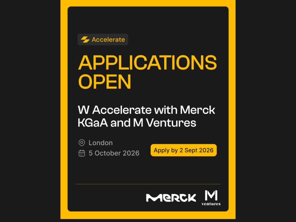
W Group has opened applications for W Accelerate with Merck KGaA and M Ventures, inviting reproductive and maternal health startups, scaleups and spinouts to pitch for direct access to global pharma partnership and strategic investment.
Selected companies will pitch on 5th October, competing for the chance to accelerate their growth through commercial partnerships, investment, or both.
This is the third time Merck KGaA, a global leader in reproductive health, has partnered with W Group on the programme, which exists to close the innovation and investment gap in women’s health by connecting the sector’s most promising startups directly with the corporates and investors positioned to scale them.
What Merck KGaA and M Ventures are looking for
This year’s call is focused on breakthrough solutions in female infertility, fertility preservation, adenomyosis, endometriosis, polyendocrine metabolic ovarian syndrome (PMOS), ovarian insufficiency, preeclampsia and pregnancy comorbidities.
New for this round, applicants choose between three pathways depending on what they need from the programme:
- The Partnership Lane, for companies seeking commercial collaborations and strategic relationships
- The Investment Lane, for founders looking to connect with investors and secure funding to scale
- The Dual Lane, for innovators pursuing both partnership and investment opportunities
How the Accelerate event works
Selected companies get a 1:1 pitch practice session ahead of time, then a private 30-minute session with Merck KGaA and M Ventures leadership on the day itself, small-group sessions with regulatory and investment strategy experts, an “Ask Merck Anything” roundtable, and a VIP networking reception.
Key dates
- Open call launches: 8th July
- Open call closes: 2nd September
- Notification of successful companies: 11th September
- Pitch day: 5th October
Applications are open now at wplatform.typeform.com/to/KGzviBQM.


 Menopause2 weeks ago
New menopause drug approved for use by NHS in Scotland

 Hormonal health1 week ago
Stardust period tracker shares health data, study reveals

 Entrepreneur5 days ago
Zero Candida market set to reach over US$2 billion by 2030 – report

 News2 weeks ago
Women’s health draws record $1.55bn in equity as capital spreads beyond the mega-rounds
 Insight2 weeks ago
Softening ovaries could extend fertility as women age, study suggests

 Entrepreneur2 weeks ago
Onto Health acquires diagnostics software company Levy Health

 News1 week ago
Congress urged to invest over $20bn to close women’s health gap

 Wellness6 days ago
Nearly 60% of young women get health advice from influencers, research finds