



Cancer
Racial disparities seen in care after abnormal mammograms


Despite similar availability of diagnostic technologies to women of different racial and ethnic groups, significant disparities were seen in who actually received same-day diagnostic services and biopsies after abnormal mammogram findings, new research shows.
Even when breast-care facilities had the necessary technology and capabilities, minority groups were less likely than white women to receive diagnostic imaging on the same day as an abnormal screening result.
Black women in particular were less likely to receive same-day biopsies, compared with white women.
“We’re looking at how the diagnostic workup process might contribute to the population disparities we see with breast-cancer outcomes,” said Dr. Marissa Lawson, assistant professor of radiology at the University of Washington School of Medicine.
“It was surprising to find similar availability (of technologies) across racial and ethnic groups. But we still saw big differences in who was getting those same-day services.”
The retrospective study spanned nearly 11 years, from January 2010 to December 2020. The data represented more than 1.1 million female patients ages 40-89 and more than 3.5 million screening mammograms. The patients were seen in six U.S. states at 136 screening facilities that, as members of the Breast Cancer Surveillance Consortium, make data available for study.
With the multilevel analysis, the researchers hoped to shed light on how factors such as race and ethnicity, household income and neighbourhood educational attainment affected access to, and use of, breast-cancer diagnostics.
In the study, they first identified which consortium facilities offered onsite access to both standard-care and advanced diagnostics and biopsy. They then measured patients’ access to these capabilities, discerned which screening mammograms yielded an abnormal result, and evaluated those patients’ receipt of diagnostic imaging and biopsy on the same day and within 90 days at facilities that had the technology and capability onsite.
The researchers adjusted for several potentially influential factors such as patient breast density, biopsy history, personal and family history of breast cancer.
Asian, Black and Hispanic/Latinx patients received fewer same-day diagnostic services after undergoing a screening mammography with abnormal results at facilities offering any diagnostic or biopsy services. Patients who lived in the lowest-income neighbourhoods were 58 per cent less likely to have same-day diagnostic evaluations than those living in the highest-income neighbourhoods.
Notably, Lawson said, rural-resident patients were more likely to receive same-day diagnostic services than were urban residents.
Black patients and those who lived in a relatively lower median income were less likely than white patients to receive same-day biopsy at a capable facility. However, Hispanic/Latinx patients were more likely than white patients to receive same-day biopsy.
“Getting a screening mammogram is an anxiety-inducing experience for a lot of people, and most patients I encounter want their results as soon as possible,” Lawson said.
“If you’re able to do a diagnostic workup on the same day, instead of making them wait to schedule a follow-up visit — which might create a headache with work or transportation — that’s better for the patient. And if treatment is advised based on imaging and biopsy, we should do all we can to shorten the time between screening and treatment (for those diagnosed with breast cancer).”
The authors acknowledged several limitations, including a study population that might not be nationally representative of women’s education and rural residency. Variables that might affect patients’ ability to take advantage to clinic services, such as employment, insurance status, transportation barriers and childcare needs, were not included in the analyses.
It also is unknown whether individual patients declined the opportunity for same-day services or were not offered such services.



GSK said its ovarian cancer drug shrank or cleared tumours in more than 60 per cent of patients in an early trial as CCO Luke Miels pushes faster development.
The company said that in an early-stage trial, Mocertatug Rezetecan, known as Mo-Rez, shrank or eliminated tumours in 62 per cent of patients with ovarian cancer after chemotherapy had failed, and in 67 per cent of those with endometrial cancer.

Hesham Abdullah, GSK’s global head of cancer research and development, said: “Treatment of gynaecological cancers remains a major challenge, with a pressing need for new therapies that offer improved response rates.
“With Mo-Rez we now have compelling evidence of a promising clinical profile.”
GSK acquired the Mo-Rez treatment, an antibody-drug conjugate, from China’s Hansoh Pharma in late 2023 and has trialled it in 224 patients around the world, including the UK, over the past year.
Only a few patients needed to stop treatment because of side effects, the most common being nausea.
It is given every three weeks by intravenous infusion, meaning directly into a vein.
Combined with data from a separate intermediate trial in China, the results have given the British drugmaker the confidence to go straight to late-stage trials, with five clinical studies planned globally in the next few months, including on patients in the UK.
Speaking to journalists before the conference, Abdullah described Mo-Rez as a “key asset” in the company’s growing cancer portfolio.
It is expected to be a blockbuster drug, with peak annual sales of more than £2bn, which GSK hopes will help it achieve its 2031 sales target of £40bn.
A few years ago GSK did not have any cancer drugs on the market, but it now has four approved medicines and 13 in clinical development.
Last year, oncology generated nearly £2bn in sales, up 43 per cent from 2024, with sales of its endometrial cancer drug Jemperli rising 89 per cent.
Article produced in association with Spital Clinic
Cervical screening saves thousands of lives from cervical cancer in the UK every year.
The programme has changed significantly in recent years, and understanding what it now involves — and what your results actually mean — is probably the most useful preparation you can do before your appointment.
Why Cervical Screening Exists — and What Changed in 2025
The NHS cervical screening programme now uses HPV primary screening — a method NHS England describes as testing for the human papillomavirus (HPV) rather than looking directly for abnormal cells.
If HPV is not found, the sample is not examined further; if HPV is found, the same sample is then checked for cell changes.
The reason HPV sits at the centre of this approach is straightforward. Nearly all cervical cancers are caused by certain high-risk types of HPV — and HPV is extremely common.
Most people encounter some form of it during their lives, and the body usually clears it without any treatment.
The problem arises when high-risk HPV persists, because over time it can cause cell changes in the cervix that, if left undetected, may eventually develop into cancer.
Cervical screening is therefore a prevention test, not a cancer diagnosis. Its purpose is to find those cell changes early, when treatment is straightforward and effective.
A significant change took effect in England on 1 July 2025.
The screening interval for those aged 25–49 was extended from three years to five years, in line with the 50–64 age group — so everyone aged 25 to 64 in England is now invited every five years.
The interval in Northern Ireland currently differs; Cancer Research UK has current details.
Who Is Invited and How to Book
NHS England invites everyone with a cervix aged 25 to 64, with the first letter usually arriving a few months before a person’s 25th birthday.
Invitations are sent automatically and are linked to GP registration — the simplest way to make sure yours arrives is to be registered with a GP and keep your contact details up to date. If your invitation hasn’t come through, any GP surgery can arrange an appointment.
Trans men and non-binary people registered as female with their GP will receive invitations automatically.
Those registered as male will not — but can self-refer through the NHS cervical screening programme website or ask their GP to arrange a test.
People over 65 are not routinely invited but are not excluded.
Anyone who has never been screened, or whose most recent result was abnormal, can request screening through their GP or a clinic.
Appointments are most commonly offered at GP surgeries, carried out by a nurse or doctor.
For those who prefer a different setting, shorter waits, or an appointment outside NHS hours, a private smear test can be arranged through specialist gynaecology clinics, often within a few days.
What Happens at a Smear Test Appointment
The test itself takes less than five minutes, with the full appointment lasting around ten minutes.
Knowing what happens — step by step — removes most of the uncertainty that makes it feel more daunting than it actually is.
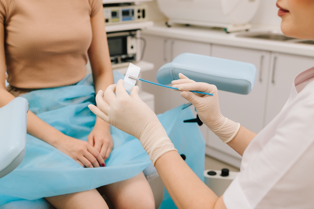
You undress from the waist down and lie on an examination table, knees bent and falling gently apart.
The nurse or doctor applies a little lubricant and gently inserts a small speculum, which is opened just enough to make the cervix visible.
A small, soft brush sweeps a cell sample from the surface of the cervix. The speculum is removed, and you get dressed.
That is the entirety of the test.
A little preparation helps both comfort and sample quality.
Avoid vaginal medicines, lubricants, and creams for at least two days beforehand, as residue can interfere with the results. Avoid scheduling during your period for the same reason.
Loose-fitting clothing — a skirt or wide-leg trousers — makes undressing and repositioning much easier.
If you are going through the menopause, vaginal dryness can make speculum insertion uncomfortable.
Cancer Research UK notes that using a short course of oestrogen cream or pessaries for around two weeks beforehand can help considerably — though stop two days before the appointment to avoid affecting the sample.
It is worth mentioning to your GP when you book.
Most people feel some pressure or mild discomfort, but it does not usually hurt.
If you find it painful, you can ask for a smaller speculum, insert it yourself, or try lying on your side — all are standard adjustments. Light spotting afterwards is normal and usually clears within a few hours. You can ask to stop at any point.
Understanding Your Results
The most common result — received by 87 in every 100 people screened — is HPV not found.
That means no high-risk HPV was detected, your risk of developing cervical cancer before the next screen is very low, and nothing further is needed until your next invitation arrives in five years.
The other results fall into two categories. Nine in every 100 people are told HPV was found, but no cell changes were detected.
This is not a cancer result and does not require immediate treatment — it means high-risk HPV is present and the cervix is being monitored.
The pathway is a repeat screen at one year; if HPV is still present, another repeat at two years. Only if it persists at that point is a colposcopy referral made.
Four in every 100 people receive an HPV-positive result alongside detected cell changes, and are referred directly for colposcopy.
That referral is not a diagnosis of cancer.
A colposcopy is a closer examination of the cervix, carried out in a clinic using a magnifying device, allowing a clinician to look in detail at any flagged cell changes.
Most people who attend colposcopy do not have cervical cancer — NHS England is clear on this. If a biopsy is taken or cells are removed, there is a small risk of bleeding and infection, both well-managed.
Those who prefer not to wait for an NHS appointment can access a private colposcopy at specialist clinics.
Results usually arrive by post or through the NHS App within two to six weeks. Samples are kept for ten years.
HPV can remain in the body for many years without symptoms, so a positive result says nothing about recent exposure or transmission history.
HPV, the Vaccine, and When to See a GP Without Waiting
Nearly all cases of cervical cancer are linked to high-risk HPV.
The virus spreads through skin-to-skin genital contact, vaginal, anal or oral sex, and sharing sex toys — it does not require penetrative sex to pass between people. The vast majority of people who carry it clear it naturally and will never know they had it.
The UK’s HPV vaccination programme, which began in 2008, has meaningfully reduced risk in younger age groups.
But vaccinated people still need to attend cervical screening — the vaccine does not protect against all high-risk HPV types. Vaccination and screening work together; one does not replace the other.
Cervical screening runs on a schedule, but some symptoms need attention straight away — do not wait for your routine invitation.
According to NHS England, these include unusual vaginal bleeding, bleeding after sex, bleeding during or after the menopause, heavier periods than usual, changes in vaginal discharge, pain during sex, or persistent lower back or pelvic pain.
These symptoms do not confirm anything, but they need investigating without delay.
Attending every invitation remains the single most important thing anyone in the eligible age group can do — and with the 2025 extension to five-year intervals, each appointment now covers a longer window than it once did.
The extension of the English screening interval to five years is backed by strong evidence about the accuracy of HPV primary screening.
Simply turning up remains, by some margin, the most protective thing anyone in the eligible age group can do.
This article is for informational purposes only and does not constitute medical advice. For personal health concerns, consult a qualified healthcare professional. Cervical screening eligibility and intervals may vary; refer to current NHS guidance or your GP for the most up-to-date information applicable to your circumstances. This piece was produced in association with Spital Clinic, which provided background clinical information for editorial purposes. Hyperlinks to external sources are included for reference only and do not represent an endorsement of any product, service or organisation.

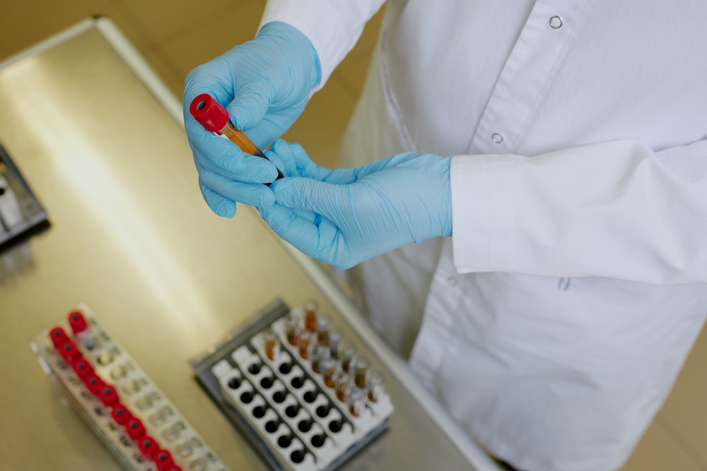
Australian startup Proseek Bio has raised AU$1.5m in an oversubscribed seed round to commercialise its ovarian cancer blood test.
With up to 80 per cent of surgeries for suspected ovarian cancer coming back benign, the company says better diagnostics are urgently needed.
Proseek Bio says its test is designed to support earlier and more precise clinical decision-making, while improving referral pathways and reducing unnecessary surgery.

OC-Triage is a blood-test-based ovarian cancer detection test that identifies specific glycoproteins, which are proteins that can indicate a future likelihood of ovarian cancer.
Proseek Bio says it could change how women are triaged for diagnostic surgery.
Founded by Michelle Hill, Proseek Bio says it has future applications beyond ovarian cancer, including endometriosis triage and other women’s health diagnostics.
The company said in a statement: “This funding will support the next stage of development for OC-Triage, our blood-based test for ovarian cancer, as we move toward clinical lab deployment and validation.”
Proseek Bio says women’s health has long been framed as “niche” or an “impact category” but says it is in fact one of the most mispriced opportunities in healthcare.
Ovarian cancer is one of the most lethal gynaecological cancers and more than 15,000 women in Australia undergo diagnostic surgery for ovarian cancer each year, with many cases diagnosed too late.
The five-year survival rate for ovarian cancer today is the same as the five-year survival rate for all cancers in 1975.
“We’re now seeing that shift: capital is beginning to catch up to the scale of both the clinical need and the commercial potential,” the company said.
“OC-Triage is designed to support earlier and more precise clinical decision-making, improving referral pathways and reducing unnecessary or delayed surgery.”
Proseek Bio says it is now preparing to pursue a Series A raise focused on market entry, clinical studies and regulatory progression.








 Entrepreneur2 weeks ago
Three sessions that show exactly where women’s health is heading in 2026

 Menopause4 weeks ago
California plans US$3.4m menopause care overhaul

 Pregnancy3 weeks ago
How NIPT has evolved and what AI NIPT means in 2026

 Hormonal health3 weeks ago
Watchdog bans five ads for women’s heath claims

 Entrepreneur4 weeks ago
WHIS USA 2026 announces first ticket release for landmark Women’s Health Innovation Summit

 Menopause4 weeks ago
Menopause has no lasting impact on cognition, research finds

 News2 weeks ago
Two weeks left to make your mark in women’s cardiovascular health

 Opinion3 weeks ago
Q1 momentum: Female founders are advancing, but the system still hasn’t caught up