


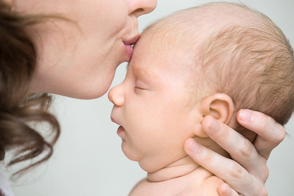
Pregnancy
Unregulated pregnancy scans putting lives in danger, experts say

High street clinics offering pregnancy scans could be putting unborn babies and their mothers in danger through a lack of properly trained staff, UK experts warn.
Hospital specialists report cases of missed health problems, misdiagnosed conditions and instances where women were wrongly told their babies had died or were malformed.
The Society of Radiographers (SoR) says high street clinics have grown rapidly in number, but anyone can buy an ultrasound machine and perform scans without qualifications.

Katie Thompson, a hospital sonographer and president of the SoR said: “I had a lady referred for a potential miscarriage from a clinic and when I scanned her they’d measured a bleed in the womb and completely missed a very early pregnancy sac with a baby inside it.”
Potentially, if they were at a private clinic that could offer a miscarriage service, they could have been given medication to bring on a miscarriage on a pregnancy that was actually not miscarrying.
The SoR says it has seen cases where private clinics wrongly diagnosed ectopic pregnancies — where the embryo implants outside the womb, which can be life-threatening — or missed actual ectopic pregnancies. Clinics have also misdiagnosed cervical problems and missed abnormalities in babies.
Elaine Brooks, a former hospital sonographer and Midlands regional officer for the SoR, said some people attended their 20-week NHS scan after having a private “sexing” scan a week or two earlier.
She said: “And then they come for their NHS scan and there’s quite a large abnormality that should have been picked up – something like spina bifida, polycystic kidneys or fluid-filled ventricles in the head – things that you wouldn’t expect to have developed in a week.”
Spina bifida is a birth defect where the spine and spinal cord do not form properly.
Polycystic kidneys involve multiple cysts in the kidneys, while fluid-filled brain ventricles can indicate hydrocephalus, a build-up of cerebrospinal fluid.
The SoR is calling for ‘sonographer’ to become a protected job title, meaning only qualified practitioners registered with a regulatory body could use it, similar to radiographer, dietitian or speech and language therapist.
Thompson said: “At the moment, absolutely anybody can go and buy an ultrasound machine and set up a practice without any qualifications whatsoever. And that has happened.
“There has been somebody that bought a machine and started scanning in her front room because after having a baby, she thought it’d be a nice thing to do.”
Thompson said it was possible for people struck off professional registers to continue offering ultrasound scans privately.
The SoR said the Health and Care Professions Council had evidence of a sonographer struck off the radiographers’ professional register for 10 years for sexual misconduct who was later employed in a private ultrasound clinic.
Thompson said the lack of a professional register made it difficult for patients to verify qualifications, but there were steps people could take.
These include checking how long a clinic has been operating, whether it is registered with and has been inspected by the Care Quality Commission, and seeking recommendations from midwives, GPs, NHS sonographers, friends or family.
“There are some excellent private clinics around that have fully qualified staff,” she said.
The Department of Health and Social Care noted that while sonography is not a legally regulated profession, sonographers can voluntarily join the Register of Clinical Technologists, allowing patients to check whether they have met professional standards.
A Department of Health and Social Care spokesperson said: “No parent should face the trauma of an incorrect diagnosis, and our sympathies are with families affected.
“We are committed to ensuring appropriate regulation for all health and care professions so patients can feel confident their care is in safe and qualified hands.
“The regulation of healthcare professionals is kept under review to ensure patient safety remains paramount.
“We will carefully consider any proposals from professional bodies regarding this.”


Women who develop pre-eclampsia face a higher risk of chronic kidney disease and high blood pressure later in life, new research suggests.
The amount of protein found in the urine during pregnancy may help identify those at greatest risk of developing long-term health problems.

Pre-eclampsia usually involves high blood pressure and increased protein in the urine. Some women also experience severe headaches and changes to their vision.
The condition cannot be treated during pregnancy and, in some cases, labour must be induced early to protect both the woman and baby.
The study found that the condition may be linked to longer-term health problems.
Anne Høy Seemann Vestergaard, a medical doctor and PhD at the department of clinical medicine at Aarhus University, said: “What we can see is a clear association between pre-eclampsia and the development of high blood pressure, chronic kidney disease and cardiovascular disease later in life.”
The researchers found that the amount of protein passed in the urine during pregnancy was linked to the risk of developing chronic conditions after giving birth.
Protein in the urine can indicate that the kidneys are not filtering blood normally.
Vestergaard said: “The most surprising finding was how clearly the amount of protein in the urine during pre-eclampsia was linked to the risk of later high blood pressure and chronic kidney disease. Women with moderate to severe protein excretion had a higher risk of both conditions compared with women with low or no protein excretion.”
Among women with pre-eclampsia and moderate to severe levels of protein in the urine, around one in 20 developed chronic kidney disease within 10 years and around one in six developed high blood pressure.
Most women in the study did not develop long-term complications, but the researchers said the increased risk should still be taken seriously because the potential effects can be severe.
Vestergaard said: “At first glance, this may sound like a low number, but it represents a markedly increased risk when the groups are compared. In the group with pre-eclampsia and high levels of protein in the urine, around 1 in 20 women developed chronic kidney disease within ten years, including early stages of the disease, compared with around 1 in 100 in the group with lower or no protein excretion.”
She added: “That is a considerable number in light of the fact that chronic kidney disease is a potentially serious condition that can progress to kidney failure if isn’t diagnosed early.”
The findings suggest women who experience pre-eclampsia may benefit from more systematic monitoring after pregnancy.
Vestergaard said: “Our study suggests that these women may benefit from monitoring of blood pressure and kidney function after pregnancy.”

Light exercise and less sitting may reduce pregnant women’s risk of serious blood pressure complications, according to a new study.
Researchers have proposed a daily activity and sleep guide that they say was linked to a nearly 30 per cent lower risk of hypertensive disorders of pregnancy.
The suggested pattern includes fewer than eight hours of sedentary time, at least seven hours of light physical activity, around 22 minutes of more intense activity and nearly nine hours of sleep.

The University of Iowa-led study examined the daily behaviours of 470 pregnant women across all stages of pregnancy.
Participants wore monitors that measured physical activity over 24-hour periods and recorded how long they spent asleep.
Hypertensive disorders of pregnancy include chronic high blood pressure, gestational hypertension and pre-eclampsia.
Gestational hypertension is high blood pressure that develops during pregnancy, while pre-eclampsia is a potentially serious condition involving high blood pressure and signs that organs may be affected.
Sedentary behaviour means being mostly inactive, such as sitting or lying down.
Light physical activity can include casual walking, moving around the home or standing.
Moderate to vigorous activity includes movement such as brisk walking, where breathing and heart rate increase.
Kara Whitaker, associate professor in the department of health, sport, and human physiology at Iowa and corresponding author of the study, said: “We are identifying the optimal composition of movement behaviours across the day associated with the lowest risk of developing HDP and the most improved health outcomes.
“This blueprint holds for each and every trimester of pregnancy.”
Study participants were enrolled at sites in Iowa City, Pittsburgh and Morgantown, West Virginia.
The women wore activity and sleep monitors for at least one week during each trimester of pregnancy.
Four in five participants were non-Hispanic white and nearly a quarter lived in rural areas.
The data showed a steep rise in risk among pregnant women who were sedentary for more than 10 hours a day.
Women who increased light physical activity to at least four hours a day reduced their risk of hypertensive disorders of pregnancy to 15 per cent from 30 per cent.
Whitaker said: “Just moving around more seems to have significant health benefits.
“And I think it also may be a more feasible target for women who are pregnant who are not exercising regularly.”
The researchers said they were surprised that longer durations of moderate to vigorous physical activity did not appear to provide additional benefit.
Sleep beyond a certain duration also did not appear to bring major further benefits.
Whitaker said: “Through this study, we are providing evidence that reducing sedentary behaviour and engaging in light physical activity are important, and maybe more important, when it comes to pregnancy and health.”
The findings may be relevant beyond pregnancy because clinical research has shown that women who develop hypertensive disorders of pregnancy are more than twice as likely to develop heart disease later in life.
Cardiovascular disease includes conditions affecting the heart and blood vessels, such as heart disease and stroke.
Whitaker said: “We know that cardiovascular disease is the number one killer of women, and if we can intervene in pregnancy and prevent women from developing a hypertensive disorder of pregnancy, we are putting them on a better trajectory, away from cardiovascular disease and toward more optimal cardiovascular health.”
The study was published online on June 10.
A second study, published online on May 27, looked more closely at the ratio and type of sedentary behaviour and light physical activity linked to a lower risk of hypertensive disorders of pregnancy.
Whitaker is a lead co-author on that study.
Co-authors in the June 10 study include Alex Crisp, Jaemyung Kim, Karina Smith, Donna Santillan, Mark Santillan and Bridget Zimmerman, from Iowa; Jacob Gallagher, from Iowa State University; Melissa Jones, from Oakland University in Michigan; Bethany Barone Gibbs, Katrina Wilhite, Alexis Thrower and Iqra Sheikh, from West Virginia University; and Sabera Rahman, Janet Catov, Christopher Kline and Maisa Feghali, from the University of Pittsburgh.
The National Institutes of Health, the University of Iowa Institute for Clinical and Translational Science, the University of Pittsburgh Clinical and Translational Science Institute and the West Virginia Clinical and Translational Science Institute funded the research.


 Menopause4 weeks ago
Perimenopause misinformation ‘putting women at risk’

 Entrepreneur3 weeks ago
Women’s Health Innovation Summit opens submissions for 2026 Innovation Showcase

 Insight2 weeks ago
British women among angriest in Europe, health survey reveals

 Menopause3 weeks ago
Women still being failed when they reach menopause, experts say

 Menopause3 weeks ago
Sweden eyes domestic production of oestrogen patches amid menopause treatment shortage

 News3 weeks ago
Three menopause innovators shortlisted for Femtech World Award

 News1 week ago
Femtech World Awards 2026: Winners revealed

 Menopause2 weeks ago
Apple Health adds menopause and perimenopause tracking