


Pregnancy
Ultrasound scans in pregnancy: A trusted guide amid emerging MRI research

For many born during the UK’s most common birth dates, their arrival often carries a festive backstory.
Christmas holidays, time off work, and the boost to sperm quality in winter—December becomes a season of new beginnings.
Fast forward nine months, and January turns into a time of fresh starts, not just for the year ahead but for expectant parents embarking on the incredible journey of pregnancy.
It’s a journey filled with hope, wonder, and, naturally, questions. Among the most important tools guiding that journey are ultrasound scans.
They’re trusted. They’re accessible. And they remain at the heart of prenatal care, even as advancements in MRI research spark curiosity.
Why Ultrasound Matters
An ultrasound scan is more than just a glimpse inside the womb.
In the NHS, two routine scans are part of every pregnancy. The dating scan, offered at around 12 weeks, sets the timeline for your pregnancy.
It confirms your due date, checks for multiple pregnancies, and ensures everything is progressing as it should.
Then there’s the anomaly scan, at 20 weeks. This one is more detailed, looking at the baby’s organs, growth, and the position of the placenta.
It identifies any structural abnormalities, providing essential information to parents and clinicians alike.
But for many parents, these NHS scans aren’t enough. They’re waiting—sometimes weeks, sometimes months—to see their baby. They want reassurance sooner.
That’s where private viability scans come in.
The Comfort of Private Viability Scans
The first trimester is a time of joy, but also of uncertainty.
It’s those early weeks, from the moment of a positive test, where questions and worries arise.
Private viability scans, available from six weeks, offer peace of mind when it’s needed most.
They confirm a baby’s heartbeat, assess gestational age, and check for multiple pregnancies.
For parents who’ve experienced loss or undergone fertility treatments, this early reassurance can be transformative.
At London Pregnancy Clinic, these scans are more than just clinical assessments.
They’re moments. Moments where parents first see their baby and begin to imagine the life they’re creating.
Performed using state-of-the-art ultrasound technology, these scans provide not only precise results but also an emotional bridge until the NHS dating scan at 12 weeks.
MRI: A Window into Complex Cases
While ultrasound is the foundation of prenatal care, MRI—magnetic resonance imaging—has carved a vital role in specific cases.
Unlike ultrasound, which uses sound waves, MRI relies on magnetic fields and radio waves to create highly detailed, cross-sectional images of the fetus and maternal anatomy.
It’s not for every pregnancy, but in complex or high-risk cases, it’s a game-changer.
Recent innovations in MRI have expanded its capabilities, allowing researchers and clinicians to see pregnancy in unprecedented detail.
MRI has proven a valuable tool for diagnosing rare fetal conditions like brain abnormalities and oesophageal atresia, especially when ultrasound results are unclear.
It is also shedding light on how pregnancy affects maternal health.
Groundbreaking studies have shown that MRI can map changes in the mother’s brain during pregnancy, offering insights into risks such as postnatal depression or conditions like migraines that may improve during gestation.
However, MRI is not a replacement for ultrasound. It is expensive, time-intensive, and less widely available, making it a complementary tool rather than a routine one.
For high-risk pregnancies or cases requiring detailed follow-up, MRI may add a level of precision that ultrasound cannot achieve alone.
The Revolution in Ultrasound Technology
Ultrasound isn’t just holding its ground—it’s leaping forward. It remains the gold standard for monitoring pregnancy because it is safe, affordable, and accessible.
And now, it’s better than ever. The GE Voluson Expert 22, for instance, represents a new era of ultrasound technology.
This isn’t just a machine. It’s a marvel.
Designed for early detection, it excels in first-trimester assessments, fetal cardiac imaging, and complex anatomy evaluations.
The Voluson Expert 22 uses advanced AI to automate key measurements. Tools like SonoLyst, SonoPelvicFloor, and SonoBiometry enhance accuracy, speed, and consistency.
For parents, this means earlier detection of potential concerns. For clinicians, it means greater confidence in every scan.
With cutting-edge 3D and 4D capabilities, this machine offers lifelike views of a baby in the womb. It’s technology that doesn’t just inform—it connects.
It allows to combine the precision of science with the compassion of care, ensuring every parent feels supported and informed.
Looking Ahead
January is a time for beginnings. For expectant parents, it’s the start of an incredible journey.
While MRI advances the science of pregnancy care, ultrasound remains the heartbeat of it all. It’s accessible, reliable, and constantly evolving.
From early viability scans to the anomaly scan at 20 weeks, these moments shape the experience of parenthood.


More than half of women with gestational diabetes report stigma from healthcare staff, family, friends and wider society, new research shows.
A survey of 1,800 UK women found widespread emotional distress at diagnosis of the condition, a form of high blood sugar that develops during pregnancy, with effects lasting beyond birth.
Gestational diabetes affects around one in 20 pregnancies in the UK, and the findings highlight the wider toll on women diagnosed with the condition.

The study was funded by Diabetes UK and led by researchers at King’s College London and University College Cork.
Dr Elizabeth Robertson, director of research and clinical at Diabetes UK, said: “Stigma can have a dangerous and devastating impact on pregnant women diagnosed with gestational diabetes, particularly at a time when emotions and anxieties may already be heightened.
“We know that stigma can lead to shame, isolation and poorer mental health, and may discourage people from attending healthcare appointments, potentially increasing the risk of serious complications.
“This research highlights the urgent need for better support systems, based on understanding and empathy to ensure no one feels blamed or judged during their pregnancy.”
More than two-thirds of women, 68 per cent, reported anxiety at diagnosis, while 58 per cent felt upset and 48 per cent experienced fear.
The psychological impact continued beyond birth, with 61 per cent saying the condition negatively affected their feelings about future pregnancies.
Nearly half of women, 49 per cent, felt judged for having gestational diabetes, while 47 per cent felt judged because of their body size.
More than 80 per cent felt other people did not understand gestational diabetes, and more than a third, 36 per cent, concealed their diagnosis from others.
Gestational diabetes stigma was also common in healthcare settings, with 48 per cent reporting that professionals made assumptions about their diet and exercise, and more than half, 52 per cent, feeling judged based on their blood glucose results.
Many women described a loss of control and a sense of disruption during pregnancy.
Nearly two-thirds, 64 per cent, felt they were denied a normal pregnancy, while 76 per cent reported a lack of control over their pregnancy.
More than a third, 36 per cent, felt abandoned by healthcare services after giving birth, and one in four, 25 per cent, continued to experience depression or anxiety postpartum.
Focus group participants described harmful stereotypes, including assumptions that they were ‘lazy’, had ‘poor eating habits’ or ‘lacked willpower’.
Comments from family and friends included remarks such as “should you be eating that?” and “you must have eaten too much, that’s why you have gestational diabetes.”
The researchers are calling for targeted interventions alongside structured emotional support for women during and after pregnancies affected by gestational diabetes, to improve both mental and physical health outcomes.
Professor Angus Forbes, lead researcher from King’s College London, said: “Stigma and emotional distress are far more common in women diagnosed with gestational diabetes than many realise.
“Everyday interactions, even with those who mean well, can deepen this harm, shaping women’s emotional wellbeing and the choices they feel able to make.
“It’s clear that meaningful action is needed to protect women’s mental and physical health.”
Risk factors for gestational diabetes include living with overweight or obesity, having a family history of type 2 diabetes, and being from a South Asian, Black or African Caribbean or Middle Eastern background.

Article produced in association with London Pregnancy Clinic
One of the most common questions in early pregnancy: NIPT or the nuchal translucency (NT) scan – do I really need both? The 2026 evidence gives a clear answer.
The two tests look at different things, and doing them together is how first-trimester screening works at its best.
This is not a debate between old and new technology. NIPT is a genuine advance in detecting chromosome abnormalities from a maternal blood sample.
The NT scan is the first detailed look at how the fetus is forming. What each sees, the other largely cannot.
What NIPT actually tells you
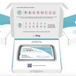
NIPT – non-invasive prenatal testing – analyses fragments of fetal DNA circulating in the mother’s blood. Taken from around 10 weeks, the test measures chromosome proportions to flag the common trisomies: trisomy 21 (Down syndrome), trisomy 18 (Edwards) and trisomy 13 (Patau).
Most panels include fetal sex and sex-chromosome aneuploidies. Extended NIPT adds selected microdeletion syndromes – most commonly 22q11.2 (DiGeorge syndrome) – and the newest whole-genome platforms can detect copy-number variants down to around 1 Mb across every chromosome.
What NIPT does not look at is anatomy. It tells you whether the chromosomes are numerically correct.
It cannot tell you how the heart, brain, spine, kidneys or abdominal wall are forming, because it analyses DNA, not structure.
The NHS offers NIPT as a second-line screening test, reserved for women who receive a higher-chance result from the combined test – precisely because NIPT is best understood as one part of a wider screening picture rather than the whole of it.
What the NT scan actually tells you
The NT scan is an ultrasound performed at 11 to 14 weeks that measures the nuchal translucency – a small fluid-filled space at the back of the fetal neck.
Protocols developed by the Fetal Medicine Foundation, the group that pioneered first-trimester screening under Professor Kypros Nicolaides at King’s College Hospital, combine the NT measurement with additional markers: nasal bone, ductus venosus flow, tricuspid regurgitation, and maternal serum biomarkers (PAPP-A and free β-hCG).
More importantly, the scan is the first structural assessment of the fetus.
Major anomalies already visible at 11-14 weeks include absence of the cranial vault, large body-wall defects such as omphalocele and gastroschisis, megacystis, severe cardiac defects with abnormal four-chamber views, and skeletal dysplasias.
An increased NT measurement itself – even with a completely normal chromosome result – is associated with a notable rate of structural heart defects and monogenic syndromes that NIPT cannot detect.
Why the combination outperforms either test alone
Taken together, NIPT and the NT scan give complementary coverage.
For the common trisomies, NIPT is more sensitive than the NT scan alone. Pooled data place detection of trisomy 21 above 99 per cent with a false-positive rate around 0.1 per cent.
Combined first-trimester screening without NIPT, using NT and serum markers alone, reaches approximately 90 per cent detection – and up to 95 per cent when nasal bone, ductus venosus and tricuspid flow are added – at a 3 to 5 per cent false-positive rate.
For that specific endpoint, NIPT is the more accurate test.
The NT scan picks up almost everything NIPT misses: structural anomalies, early markers of monogenic syndromes, confirmation of viability, accurate dating, twin chorionicity, and placental position.
An increased NT with a normal NIPT result shifts the clinical conversation toward syndromes like Noonan, Kabuki and the skeletal dysplasias – conditions with single-gene origins rather than chromosomal ones.
Working out which is which often requires genetic testing beyond NIPT. Carrier screening and expanded genetic panels – including those offered at Jeen Health – cover the single-gene territory that NIPT does not address.
When the combination matters most
Several patient groups have most to gain from doing both:
- Women conceiving after IVF or with donor gametes, where maternal age and fertility treatment each subtly shift risk profiles
- Women aged 35 and over, where baseline chromosomal risk is higher and soft markers are more likely
- Anyone with a previous pregnancy affected by an anomaly or loss, where reassurance matters
- Twin pregnancies, where NIPT performance depends on fetal fraction and structural assessment is more complex
- Women who have had a raised or borderline result on earlier screening markers
Chromosomes and anatomy are two separate clinical questions. Each needs its own answer.
What happens if the tests disagree
Disagreements between NIPT and the NT scan are not failures of either test – they are the reason both are done.
- NIPT low-risk, NT raised: consider monogenic syndromes, structural cardiac assessment, and early anomaly ultrasound follow-up
- NIPT higher-chance, scan normal: confirmatory diagnostic testing (CVS or amniocentesis) before any major decision
- NIPT no-call: repeat sampling, gestational age check and clinical review – a no-call itself is associated with an increased chromosomal risk
- Both abnormal: a clear indication for specialist fetal medicine review and early diagnostic testing
Professional guidance from the RCOG supports this complementary approach, emphasising that NIPT is a screening rather than a diagnostic test, and that its results are most useful when interpreted alongside ultrasound findings.
Practical guidance for 2026
The most efficient way to run both tests is in a single appointment window, between 10 and 14 weeks, with the blood sample taken first and the scan performed on the same visit.
Results typically return within 5 to 10 working days for standard NIPT panels, and same-day for the scan itself.
This is the logic behind the SMART Test at London Pregnancy Clinic – extended NIPT paired with a full first-trimester ultrasound in a single appointment, delivering both chromosomal and structural information in one visit. For most patients, it removes the false choice of picking one over the other.
The wider picture
The question of NIPT versus NT scan has a settled clinical answer in 2026: the two tests examine different aspects of the pregnancy, and the most complete first-trimester assessment uses both.
For a pregnancy a woman wants to carry with the fullest possible picture, both tests belong in the first-trimester window. The question worth asking is which clinic offers them together, with the pre- and post-test care that makes the results usable.
If you are deciding on first-trimester screening, a consultation with a fetal medicine specialist is the most useful first step.
Disclaimer: This article is produced for informational purposes only and does not constitute medical advice, diagnosis or treatment. Clinical guidance referenced reflects published NHS, Fetal Medicine Foundation and RCOG standards as at April 2026. Individual circumstances vary; readers are advised to consult a qualified healthcare professional before acting on any information in this article. This piece was produced in association with London Pregnancy Clinic, which provided background clinical information for editorial purposes. Hyperlinks to external sources are included for reference only and do not represent an endorsement of any product, service or organisation.


 Entrepreneur3 weeks ago
Three sessions that show exactly where women’s health is heading in 2026

 Pregnancy3 weeks ago
How NIPT has evolved and what AI NIPT means in 2026

 Menopause4 weeks ago
Watchdog bans five ads for women’s heath claims

 Entrepreneur4 weeks ago
WHIS USA 2026 announces first ticket release for landmark Women’s Health Innovation Summit

 Menopause4 weeks ago
Menopause has no lasting impact on cognition, research finds

 News3 weeks ago
Two weeks left to make your mark in women’s cardiovascular health

 Opinion3 weeks ago
Q1 momentum: Female founders are advancing, but the system still hasn’t caught up

 News4 weeks ago
Endometriosis firm wins NIH prize