


Cancer
Common cholesterol drug shows ovarian cancer promise

A common cholesterol drug could help weaken a fluid shield that helps ovarian cancer tumours survive, early lab findings suggest.
The findings do not show the drug treats ovarian cancer. But they suggest changing the environment the cancer depends on could make it more vulnerable to existing treatment.

A federally funded study at Duke University School of Medicine found that ascites, a build-up of fluid in the abdomen, may do more than cause discomfort.
Doctors can drain ascites to ease pain, improve mobility and make breathing easier, but the fluid may also help cancer cells survive and spread. It occurs in 90 per cent of people with advanced ovarian cancer.
According to the study, ascites acts as a shield, helping cancer cells evade ferroptosis, a form of cell death.
Ferroptosis is a kind of cellular rusting. It happens when iron inside a cell reacts with certain fats, causing the cell membrane to break apart.
Many metastatic cancer cells, meaning cells that float freely through the abdomen looking for new places to grow, are naturally vulnerable to this kind of damage.
“Doctors have mostly viewed ascites as a symptom rather than an active driver of disease,” said Jen-Tsan Chi, professor in the department of molecular genetics and microbiology and co-leader of the Cancer Biology Program at the Duke Cancer Institute.
“We’ve learned it gives cancer a survival advantage, which fills a major gap in understanding how ovarian cancer spreads.”
Scientists bathed cancer cell lines and patient-derived tumour cells in ascites collected from patients and watched how they responded to ferroptosis triggers.
The fluid protected cancer cells by changing how they store fats and control iron levels, effectively blocking cell death.
The protection required only trace amounts, with as little as 2 per cent immersion shielding cancer cells from destruction.
“What surprised us was how selective this effect was,” said Yasaman Setayeshpour, first author and graduate student in molecular genetics and microbiology at Duke School of Medicine.
“Ascites didn’t protect the cancer cells from other well-known types of cell death, like apoptosis or necrosis, it only blocked ferroptosis.
“To figure out why, we broke ascites down into major parts, like lipids, proteins, and small molecules, and tested what happened when each was removed.
“When we took the lipids out, the protective effect disappeared. That told us lipids are the key reason ascites helps these cancer cells survive.”
But researchers found an unexpected helper in bezafibrate, an older cholesterol drug used to lower triglycerides by altering how the body processes fats.
The cholesterol drug restored sensitivity to ferroptosis, but only when ascites was present. On its own, the drug did not trigger cell death or slow tumour growth in mice.
The drug’s impact depended on the cancer’s surroundings, in this case the fat-rich fluid bathing the tumour. Researchers found that targeting this environment, using repurposed drugs like bezafibrate, could leave cancer cells more exposed to existing cancer treatments.
Chi said the finding could have implications beyond ovarian cancer. Other cancers, including colorectal and pancreatic cancers, can also spread within the abdominal cavity.
“This work shows how much the environment around a tumour matters,” Chi said.
“Biological fluids like ascites don’t just give cancer cells a place to move. They actively help drive how cancer spreads.”


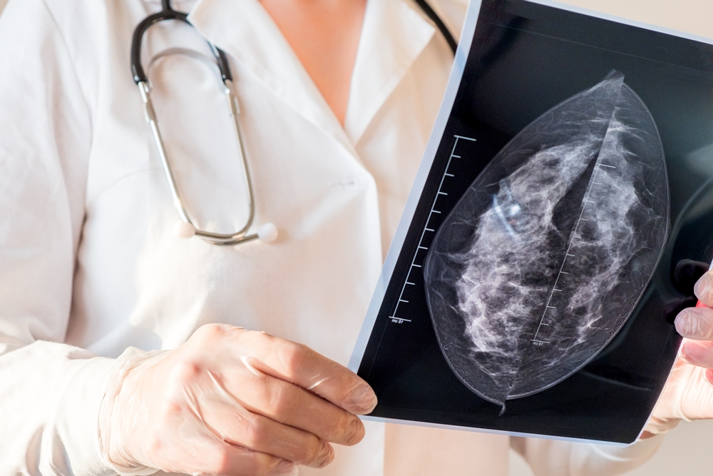
Changes in AI mammogram scores may help predict breast cancer years before diagnosis, research involving more than 54,000 women suggests.
Scores rose steadily among women who later developed the disease but remained broadly stable among those who did not.
The increase could be detected up to six years before diagnosis and became much steeper during the final two years.

Researchers led by Professor Constance Lehman, of Harvard Medical School and healthcare technology company Clairity, analysed screening mammograms taken between 2009 and 2019.
They used a validated, open-source deep learning model to calculate five-year breast cancer risk scores from the images alone.
Deep learning is a form of artificial intelligence trained to recognise complex patterns in large amounts of data.
The model examined the whole mammogram rather than relying on a limited, predetermined feature such as breast density.
Models of this kind have performed better than traditional risk models and breast density alone when estimating a woman’s five-year breast cancer risk.
The study initially included 239,703 consecutive two-dimensional screening mammograms from 89,882 patients across six imaging sites spanning urban tertiary, community-based and rural settings.
All were standard bilateral full-field digital mammography examinations, taken with or without digital breast tomosynthesis.
Digital breast tomosynthesis uses multiple low-dose X-ray images to create a three-dimensional view of the breast.
After exclusions, the final analysis involved 54,014 women with a median age of 61 and a total of 158,807 mammograms.
Each woman contributed one index examination and up to six previous annual mammograms. Women had a median of three scans each.
For women who developed cancer, the index examination was their final screening mammogram within the year before diagnosis. For the cancer-free group, it was their final mammogram during the five-year study period.
The model did not use demographic information, clinical records or historical imaging data when calculating each score.
Of the women included, 817, or one per cent, were diagnosed with breast cancer within 365 days of their index examination.
This included 451 women, or 55 per cent, with invasive breast cancer and 118, or 14 per cent, with ductal carcinoma in situ, known as DCIS.
DCIS occurs when abnormal cells are found inside a milk duct but have not spread into the surrounding breast tissue.
The cancer type was unknown for the remaining 248 patients, representing 30 per cent of the cancer group.
A total of 682 cancers, or 83 per cent, were detected through screening, while 135, or 17 per cent, were interval cancers diagnosed between routine mammograms.
The other 53,197 women were not diagnosed with breast cancer during follow-up and formed the cancer-free comparison group.
Professor Lehman said: “We observed clinically relevant differences in risk trajectories between women who did and did not develop cancer. The increase in scores among cancer patients was detectable as early as six years prior to diagnosis and became more pronounced over time.”
Among women later diagnosed with the disease, the median score rose from 2.1 five to six years before diagnosis to 6.6 at the index examination.
Scores among cancer-free women remained stable, with median values ranging from 1.8 to 2.2 throughout the study.
The rise among women who developed cancer was steepest during the two years before their index examination.
Professor Lehman said: “These findings demonstrate signals, invisible to the human eye, in the image alone can predict future risk. This is exciting, because 85 per cent of women diagnosed with breast cancer do not have a significant family history of breast cancer or known genetic mutations.”
Most breast cancers are considered sporadic, meaning they are not driven by inherited genetic changes or a family history of the disease.
Traditional risk models have a limited ability to distinguish between women who will and will not develop breast cancer when used across large screening populations.
Researchers said tracking how scores change over time could provide more information than calculating risk at a single appointment.
Professor Lehman said: “AI-derived risk scores can identify patients who are otherwise predisposed to the disease, and our findings demonstrate that image-based AI risk scores evolve over time and that changes in those scores may provide additional information about future breast cancer risk.”
The patterns remained consistent when women were grouped by age and breast density.
Breast density describes the amount of fibrous and glandular tissue visible on a mammogram. Dense tissue can make cancers harder to detect and is also associated with an increased risk of the disease.
Researchers said image-based scores could support personalised screening and risk-reduction strategies without relying on self-reported or inconsistent clinical information.
Professor Lehman said: “These trends remained robust across subgroups defined by age and breast density, further supporting the generalisability of our findings. This is particularly relevant given persistent disparities in screening performance across patient populations. A dynamic biomarker approach grounded in the imaging data could mitigate some of these disparities by enabling risk-based personalisation that does not rely on self-reported or inconsistent clinical data.”
A biomarker is a measurable sign that can indicate a person’s health, disease risk or response to treatment.
Changing scores could eventually help clinicians identify women who may benefit from additional imaging or measures intended to reduce their risk.
Professor Lehman said: “With the power of AI, computer vision, and the ability to extract predictive data, we are able to apply the power of imaging to risk assessment and preventing disease from developing. Having a dynamic risk score opens up a whole new domain of more effective preventive therapies for breast cancer, similar to how we screen for and treat patients with high cholesterol and hypertension.”
AI image-based risk scores are included in the 2026 National Comprehensive Cancer Network guidelines.
The guidelines recommend that, from the age of 35, women with an elevated five-year risk score of more than 1.7 per cent consider breast MRI alongside annual mammography.
An AI image-based model approved by the US Food and Drug Administration is already being used to calculate five-year breast cancer risk at selected US healthcare institutions.

WID-easy, the only non-invasive triage test for endometrial cancer in routine use in a European public health system, has been cited in Germany’s highest-tier clinical guidance; a marker that non-invasive detection is reaching clinical maturity.
The vaginal-swab test designed to spare women unnecessary invasive procedures has been referenced in the updated German S3 Guideline on Endometrial Cancer, its maker Sola Diagnostics has announced.
The Austria-based women’s-health diagnostics company behind the WID-easy Test, said the test now features in the recommendations-supporting background text of the guideline’s latest version (v4.0, May 2026; AWMF 032-034OL), in Section 4.3.
The S3 designation is the highest evidence- and consensus-based tier in the German clinical guideline system, broadly comparable in standing to NICE guidance in the UK, and is widely drawn on in clinical practice, reimbursement and liability assessments.
In Section 4.3, the guideline cites four peer-reviewed validation studies of the WID-easy Test and credits it with a sensitivity of more than 95 per cent and a negative predictive value of at least 99.7 per cent.
It describes a fall in invasive workup from 19 to two dilatation-and-curettage (D&C) procedures per cancer detected when compared with transvaginal ultrasound alone, assuming a realistic 3.4 per cent cancer prevalence in women with postmenopausal bleeding, and states that the test has the potential to improve the diagnostic workup of women with peri- and post-menopausal bleeding by cutting the rate of invasive procedures.
A growing burden, an imperfect standard
Endometrial cancer is the most common gynaecological cancer in high-income countries, and its incidence is rising, driven by ageing populations and increasing obesity, making it one of the fastest-growing cancer burdens in women’s health.
A number of groups are now developing non-invasive tests for earlier detection. The current standard, transvaginal ultrasound, is an imperfect triage tool: it misses serous carcinomas and performs especially poorly in black women, a group with disproportionately high endometrial-cancer mortality.
WID-easy has been validated prospectively across multiple cohorts, including a dedicated cohort of black women in Ghana (Ken-Amoah et al., 2025).
Adopted in routine care
WID-easy is the only endometrial-cancer triage test in Europe with real-world adoption in a public health system. It is UKCA-marked and in use across NHS pilot sites in England and Scotland, and is delivered through commercial laboratory partners across the DACH region of Austria, Germany and Switzerland. Its UK pivotal study, EASY-CARE, is funded by a competitively awarded NIHR i4i grant.
The postmenopausal bleeding pathway has been singled out for change across three UK Government strategy documents published in 2026 — the National Cancer Plan for England (DHSC), the renewed Women’s Health Strategy for England (DHSC) and the National HealthTech Access Programme (NICE).
The NICE initiative names speeding up access to better tools for detecting endometrial cancer in women with unexplained bleeding as one of only four priority areas.
WID-easy is the only non-invasive endometrial-cancer triage test that is UKCA-marked and commercially available for NHS use today, with no competing molecular test yet on the market.
“Seeing WID-easy referenced in a guideline of this standing confirms that the science behind non-invasive endometrial cancer detection has reached clinical maturity,” said Prof Martin Widschwendter, founder and member of the scientific advisory board at Sola Diagnostics.
“Our goal has always been to spare women unnecessary invasive procedures without missing the cancers that matter — and to do so equitably, across all populations.”
The WID-easy Test detects and triages endometrial cancer from a vaginal swab using DNA methylation. It is built on the WID-qEC biomarker, exclusively licensed from University College London Business and complemented by Sola’s own patent portfolio. The same methylation platform underpins a pipeline of further tests in cervical, ovarian and breast cancer.

The HPV vaccine has cut the risk of dying from cervical cancer before age 30 to almost zero among those vaccinated at 12 or 13, research suggests.
The first study of its kind found that deaths have fallen sharply since school-age girls began being offered the jab in 2008, with around 200 lives saved in England so far.
Between 2020 and 2024, no cervical cancer deaths were recorded among women aged 20 to 24, the first time this had happened over a five-year period.
Without vaccination, around 23 deaths would have been expected.
Professor Peter Sasieni, lead researcher at Queen Mary University of London, said: “It’s incredible to think that a single jab can almost eliminate a particular type of cancer.”
Around 3,300 people are diagnosed with cervical cancer each year in the UK, making it the 14th most common cancer among women.
Human papillomavirus, or HPV, is thought to cause 99 per cent of cervical cancer cases. The virus is spread through close skin-to-skin contact.
Most HPV infections clear up without causing problems, but some can cause abnormal cell changes that may lead to cancer years later.
The study’s authors expect deaths from cervical cancer to continue falling as more people receive the jab and vaccinated generations grow older.
Cancer Research UK, which funded the research, called the findings an “incredible milestone” but warned that vaccination rates in England remained below recommended levels.
Michelle Mitchell, chief executive of Cancer Research UK, said: “We know the HPV vaccine is extremely effective at stopping cervical cancer before it starts and for the first time these findings show it is saving lives.”
Professor Sasieni, who specialises in cancer epidemiology at Queen Mary University of London, described the reduction in deaths since the vaccine was introduced as the “tip of the iceberg”.
Cancer epidemiology examines patterns of cancer and how the disease affects different groups of people.
He said: “As vaccinated generations grow older, we’ll see many more lives saved from cervical cancer.
“New research shows just how vital it is to keep HPV vaccination levels high so more people are protected.”
The UK government has pledged to eliminate cervical cancer as a public health problem by 2040.
However, the latest figures show vaccination rates have fallen below recommended levels.
UK Health Security Agency data shows that 76 per cent of girls in England were vaccinated by the age of 15 in 2024-25, well below the 90 per cent level the World Health Organization says is needed to eliminate cervical cancer.
Mitchell said: “It’s essential that the UK Government and health systems urgently address this with targeted action to reach communities where uptake is the lowest.”


 Insight3 weeks ago
British women among angriest in Europe, health survey reveals

 News4 weeks ago
Women still being failed when they reach menopause, experts say

 Menopause3 weeks ago
Apple Health adds menopause and perimenopause tracking

 News2 weeks ago
Femtech World Awards 2026: Winners revealed

 News4 weeks ago
Three menopause innovators shortlisted for Femtech World Award

 Menopause3 weeks ago
Menopause workplace toolkit launched to help UK employers support staff

 News3 weeks ago
Elation Health acquires EHR startup Aster

 News3 weeks ago
Endometriosis documentary profiles stars including Marilyn Monroe and Amy Schumer