




To receive the Femtech World newsletter, sign up here.
Pregnancy
AI technology could detect pregnancy-related heart disease
A digital stethoscope has been shown to identify twice as many cases of peripartum cardiomyopathy as compared to regular care

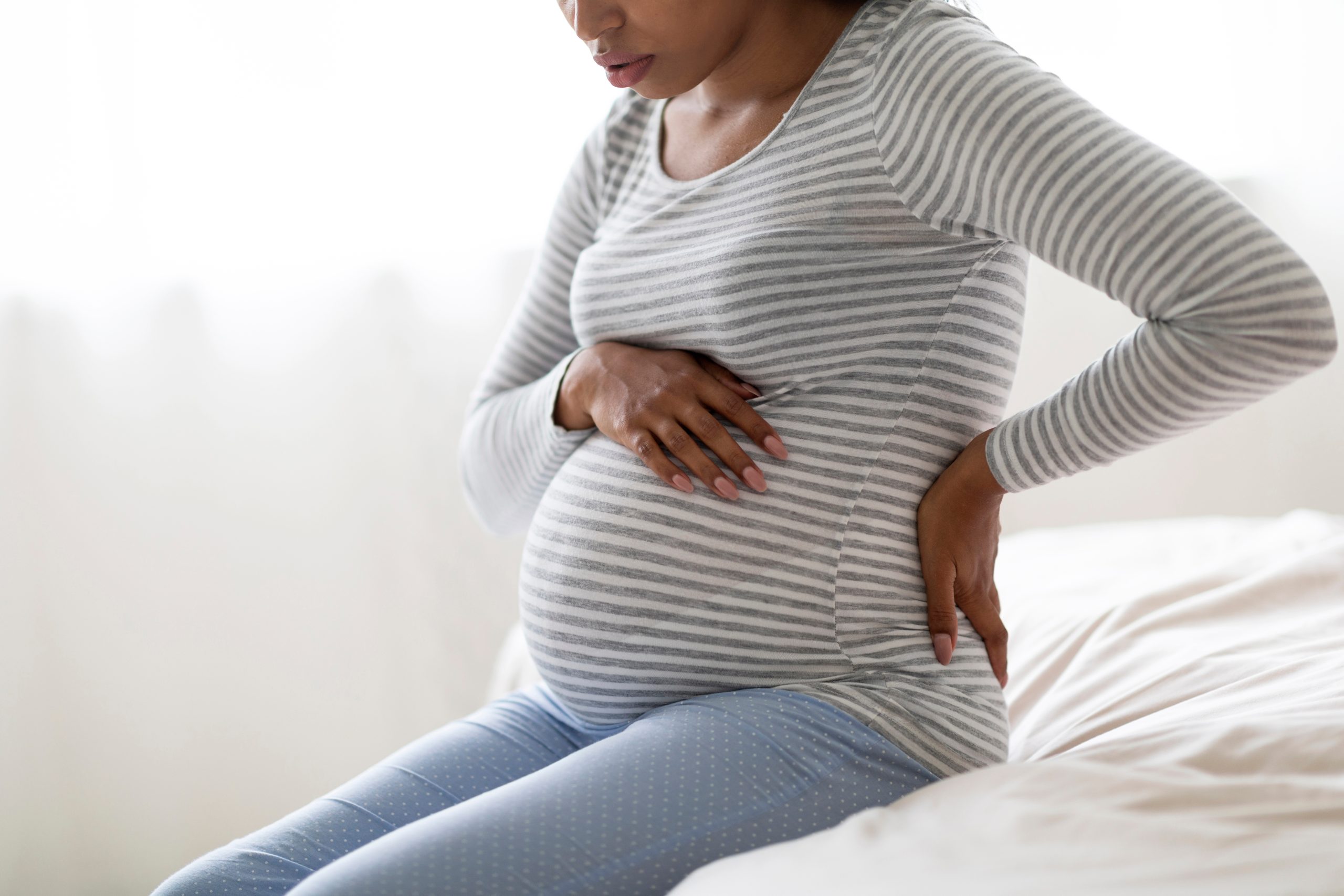
An AI-enabled digital stethoscope could detect pregnancy-related heart disease, new research has shown.
The study, conducted by Mayo Clinic, has found that an AI-enabled digital stethoscope could improve the diagnosis of peripartum cardiomyopathy, a potentially life-threatening and treatable condition that weakens the heart muscle of women during pregnancy or in the months after giving birth.
Researchers used the AI-enabled digital stethoscope that captures electrocardiogram (EKG) data and heart sounds to identify twice as many cases of peripartum cardiomyopathy as compared to regular care.
“We demonstrated for the first time in an obstetric population that AI-guided screening using a digital stethoscope improved the diagnosis of this potentially life-threatening and treatable condition,” explained lead study author Demilade A. Adedinsewo, an assistant professor of medicine in the department of cardiovascular medicine at Mayo Clinic in Jacksonville, Florida.
“This research can change current clinical practice from one that is reactive and symptom-driven to a more proactive approach of identifying pregnancy-related cardiac dysfunction using a simple, low-cost and effective screening tool.
“Earlier diagnosis would facilitate prompt and appropriate management of cardiomyopathy and reduce associated disease and death.”
Peripartum cardiomyopathy is a type of heart failure that can affect women late in pregnancy or after pregnancy. The disease weakens the heart, resulting in a decrease in the amount of blood that is pumped from the heart to other parts of the body.
It is typically diagnosed towards the end of pregnancy or in the months following and may be difficult for health professionals to detect because many of the symptoms are similar to those seen with normal pregnancy, such as shortness of breath and swelling in the feet and legs, according to the American Heart Association.
The rate of peripartum cardiomyopathy is somewhat low in the US, affecting one in every 1,000 to 4,000 pregnancies. However, it is more prevalent in countries like Nigeria, which has the highest reported incidence of peripartum cardiomyopathy worldwide, with it impacting as many as one in 96 pregnancies.
As part of the study, researchers from Mayo Clinic looked at almost 1,200 Nigerian women who were pregnant or had recently had a baby.
Approximately half were evaluated with AI-guided screening using the digital stethoscope and half received usual obstetric care in addition to a clinical EKG. Peripartum cardiomyopathy was detected twice as often among study participants when EKG testing was performed with a digital stethoscope using an AI algorithm, compared to clinical EKG in addition to routine obstetric care.
Overall, four per cent of the pregnant and postpartum women in the intervention arm of the clinical trial had cardiomyopathy compared to two per cent in the control arm, suggesting that half are likely undetected with usual care.
“While we expected AI-guided screening to improve the diagnosis of cardiomyopathy, we did not anticipate the frequency of cardiomyopathy diagnosis would be doubled,” Adedinsewo said.
She added: “Additional large trials enrolling a diverse group of women in other geographic locations are needed to evaluate the impact of AI-guided screening on cardiomyopathy diagnosis as well as its impact on adverse maternal outcomes.”



Paracetamol use in pregnancy was not linked to autism or ADHD, while separate research found reproductive differences in girls exposed before birth.
One study analysed health records from more than 120,000 children and found no increased risk of autism following prenatal paracetamol exposure.
A separate analysis of nearly 100,000 children also found no increased risk of ADHD among those born to mothers who used the painkiller during pregnancy.

Researchers from the Hong Kong Hospital Authority examined electronic health records covering pregnancies between January 2001 and December 2023.
The autism analysis included 124,333 children, who were nine years old on average and split almost evenly between males and females. There were 3,445 autism diagnoses, representing 2.8 per cent of the group.
The ADHD analysis involved 97,285 children, who were seven years old on average and also split evenly between males and females. There were 5,168 ADHD diagnoses, representing 5.3 per cent.
Women prescribed paracetamol during pregnancy were more likely to be older and have pre-existing conditions including psychiatric disorders, as well as reasons for taking the drug such as infection, fever or chronic pain.
No association was found between prenatal paracetamol exposure and either autism or ADHD.
The findings did not differ according to the trimester in which paracetamol was taken or whether use was intermittent or daily. Advanced maternal age, defined as pregnancy in women over 35, did not alter the findings.
The researchers wrote: “Paracetamol remains a safe and essential analgesic [pain reliever] and antipyretic [fever reducer] during pregnancy, whereas alternatives, such as NSAIDs and opioids carry well-documented risks.
“Unwarranted reluctance to use paracetamol could lead to undertreatment of pain and fever, or the use of more harmful alternatives, both posing risks to the pregnancy and developing fetus.”
The authors said women should assess paracetamol use with guidance from their doctor.
A separate study involving 685 pregnant women without pre-existing conditions and 302 infant daughters found associations between prenatal paracetamol exposure and differences in reproductive organs and hormone levels.
Researchers from Copenhagen University Hospital enrolled the women during their first trimester and assessed them during the first trimester, third trimester and again when their babies were three months old.
At around three months, infants experience a temporary rise in reproductive hormones sometimes called mini-puberty.
Pregnant participants completed questionnaires every two weeks about their use of pain medicines including paracetamol. Infant girls underwent ultrasound scans of their reproductive organs and blood tests to measure hormone levels.
Researchers also examined a separate group of 1,210 girls followed from infancy to adolescence whose mothers reported paracetamol use during the third trimester.
Three-month-old girls exposed to paracetamol before birth had an average 40 per cent smaller ovarian volume, 13 per cent smaller uterine volume and 23 per cent fewer ovarian follicles.
Girls exposed during the first trimester also had lower levels of Anti-Müllerian hormone, a marker of ovarian function.
Among the older girls, those exposed before birth were more likely to have smaller uteruses at puberty and smaller ovaries during their teenage years.
Dr Margit Bistrup Fischer, lead study author and postdoctoral researcher in the Department of Growth and Reproduction at Rigshospitalet hospital in Denmark, said: “Animal studies have demonstrated that impaired formation of ovarian follicles can lead to reduced fertility and earlier reproductive aging.
“Whether the differences observed in our study have implications for fertility and age at menopause in humans remains unknown and will require long-term follow-up of the girls in our cohort.”
She cautioned that women who had used paracetamol during pregnancy “should not be alarmed by our findings”, as the study found associations rather than direct causation and outcomes for individual women and children are unclear.
“Importantly, our study does not evaluate whether [acetaminophen] causes reproductive problems, nor does it provide evidence that prenatal exposure affects future fertility or age at menopause,” she said.
“Although we observed similar associations in an independent cohort, long-term follow-up is needed to determine whether these early-life differences have any clinical significance later in life.”

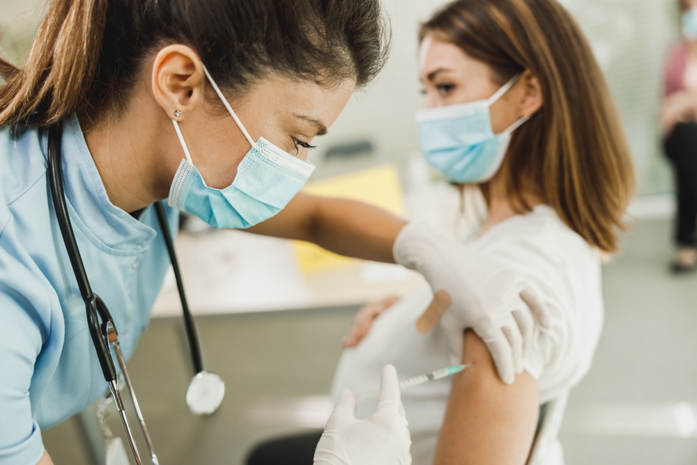
Pregnant women are among the first groups offered the NHS flu vaccine this winter, which has been updated to better match the subclade K strain.
Schoolchildren are also being prioritised and, like pregnant women, can receive the vaccine from September because they can catch and spread flu easily.
Protecting them also helps others, including younger siblings and grandparents.
Other eligible groups, including people aged 65 and over, will be able to receive a free NHS flu vaccine from October and should receive an invitation by phone or post.
Dr Amanda Doyle from NHS England said: “Last year’s flu season came early and was made worse by a new variant, which had a major impact for patients and put enormous pressure on urgent and emergency care services across the country.
“With the NHS experiencing its busiest summer on record, staff have started preparing for this winter earlier than ever – and with services already under significant pressure, it is vital that everyone offered the vaccine comes forward.”
Last winter’s flu season began more than a month earlier than usual as a new mutated strain circulated.
The strain was sometimes referred to as “Super flu”, although this is not a medical term and does not mean the virus is harder to treat. The name reflected that people had yet to build up immunity against it.
Flu vaccines are updated each year to better match the influenza viruses expected to circulate. These viruses continually mutate, so experts must predict how they are likely to change.
This winter’s vaccine includes changes intended to provide a better match for subclade K.
It remains unclear when flu will begin circulating widely this winter or how severe the season will be. Experts often look at flu activity in southern hemisphere countries such as Australia and New Zealand for indications of how the UK season could develop.
Their flu season peaks in July or August. Australia has recorded fewer cases than last year, while New Zealand has experienced high numbers.
Everyone is at risk of catching flu, regardless of age or how fit and healthy they are.
Doyle said: “Flu can really knock you for six, and for some children and pregnant women it can mean becoming seriously ill or ending up in hospital.”
The flu vaccine cannot give you flu. Instead, it helps the body know how to fight the virus.
Children aged two and over receive a nasal spray version, while adults receive an injection. One dose is needed ahead of winter.
Free NHS flu vaccines are available through pharmacies, GPs and schools. People who are not eligible for a free vaccination can pay around £10 to £20.
Schoolchildren and pregnant women can receive a free NHS flu vaccine from September onwards.
From October, free vaccination will also be available to children aged two or older who have not yet started school, people aged 65 and over, those with certain long-term health conditions, care home residents and carers of older or disabled people, including those receiving a carer’s allowance.
It will also be available to people living with someone who has a weakened immune system and those experiencing homelessness or staying in a homeless hostel or night shelter.
More adults will also be offered vaccination against respiratory syncytial virus (RSV), a virus that attacks the lungs, this winter.
From September, adults with certain pre-existing health conditions, such as chronic lung disease, will be invited for the RSV vaccine when they turn 65 rather than waiting until 75.








 Entrepreneur1 week ago
KOVE Medical raises €1.7 million to improve safety of foetal surgery

 Pregnancy1 week ago
UK study aims to transform maternity care for high-risk pregnancies

 Mental health2 weeks ago
PMDD after SSRIs or hormones: Why the brain may be the missing treatment target
 Diagnosis2 weeks ago
Ark Surgical secures further institutional backing to accelerate US expansion

 Entrepreneur3 days ago
Last chance to save on Women’s Health Week and Women’s Sport Summit: Early Bird pricing ends 11 September

 Menopause6 days ago
Menopause hormone treatment may ease brain fog, study suggests

 Motherhood2 weeks ago
Thousands of UK women develop undiagnosed PTSD after childbirth each year – study
 Cancer2 weeks ago
AI analysis of mammograms can detect heart disease, study suggests
23 Comments