


Why the first trimester is women’s health’s most underused screening window
Posted in partnership with London Pregnancy Clinic
The most detailed look most parents in England get at their developing baby arrives around the midpoint of pregnancy.
The NHS Fetal Anomaly Screening Programme offers a screening scan between 18 and 20 weeks, and that scan screens for eleven physical conditions. It is a well-run, well-audited programme, and for the overwhelming majority of pregnancies it does precisely what it was designed to do.
It was also designed around what ultrasound could reliably achieve at the time the pathway was set. The equipment has moved on considerably since. The timetable has not.
That gap – between what the pathway assumes about imaging and what imaging can now actually deliver – is one of the more interesting unexploited openings in women’s health. The sector has been noticeably slow to argue for it.
A note on vantage point. At London Pregnancy Clinic, detailed structural assessment at 10 weeks is routine rather than exceptional, and the argument below draws on that caseload rather than on theory.
The screening calendar is a product of its era
The architecture of English antenatal screening is sound. First-trimester combined screening for trisomies is offered from 11 weeks and 2 days to 14 weeks and 1 day.
The anomaly scan follows at 18 to 20 weeks. Both windows exist for good reasons, and both are set out in the NHS Fetal Anomaly Screening Programme handbook.
But note what the first-trimester contact is principally for. In the national pathway, it is a chromosomal screening appointment with a dating function attached.
The ultrasound is there largely to measure nuchal translucency and confirm gestational age. Structural assessment is, by design, deferred to the second trimester.
That division of labour made sense when first-trimester imaging could not resolve fetal anatomy in any meaningful detail. It makes considerably less sense now.
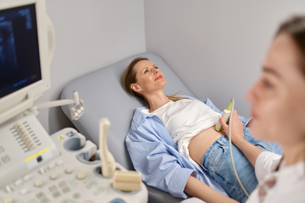
Modern high-frequency transvaginal probes and improved 3D reconstruction can visualise the fetal brain, heart, abdominal wall, limbs and spine far earlier than the pathway anticipates.
A proportion of major structural anomalies – some of them among the most consequential a family can face – are identifiable well before 14 weeks.
What earlier screening does and does not offer
Precision matters here, and overclaiming would be both wrong and counterproductive.
Earlier scanning does not replace the 20-week scan. Some anomalies are simply not present or not visible in the first trimester; certain cardiac, renal and cerebral findings declare themselves only as gestation advances.
Any serious proposition for earlier screening is additive, not substitutive. Clinics that imply otherwise do the field no favours.
What earlier scanning offers is time.
A family who receives a significant finding at 11 weeks has a materially different set of options from one who receives the same finding at 20 weeks – more room for specialist referral, for diagnostic testing, for second opinions, for genetic counselling, and for decisions taken without the compression of a looming legal and clinical deadline.
The clinical information may be the same. The circumstances in which it is received are not remotely the same.
There is also a reassurance argument that the sector tends to undervalue because it is difficult to put on a spreadsheet.
The stretch between a positive test and the mid-pregnancy scan is, for many women, a long and anxious one – and particularly so for those with a previous loss, a prior affected pregnancy, or a known family history.
That anxiety has measurable downstream costs in unscheduled contacts, primary care appointments and early pregnancy unit attendances.
The constraint is workforce, not physics
If the technology permits earlier detailed imaging, why has the pathway not moved?
The honest answer is capacity. Detailed first-trimester anatomical assessment is operator-dependent to a degree that second-trimester scanning is not.
It demands more time per appointment, more specialised equipment and, above all, sonographers and fetal medicine clinicians trained specifically in early anatomy.
Those people are in short supply across the NHS, and the training pipeline to produce more of them is not currently sized for a national shift.
A screening programme cannot responsibly offer something it cannot deliver uniformly.
Variable quality in early anatomical screening would be actively harmful: false reassurance in one unit and unnecessary alarm in another is worse than a consistent, later scan. The caution is justified.
But “this cannot be delivered everywhere tomorrow” is a different proposition from “this should not be a strategic priority”.
At present, early structural assessment sits almost entirely in the independent sector and a handful of specialist units – which makes it, in practice, a service available to those who know it exists and can pay for it. That is an equity problem the sector should be naming more plainly than it does.
Where the independent sector fits
Private clinics working at this end of the pathway have an obligation that goes beyond selling appointments.
They hold the practical experience of what early anatomical screening actually involves at volume – which views are obtainable at which gestations, where the failure modes are, how long an appointment genuinely takes, how findings should be communicated when diagnostic certainty is still some weeks away.
That is evidence the wider system needs, and the honest route for an independent provider is to generate it, publish it and contribute it to the bodies that set standards rather than hold it as a commercial advantage.
The operational questions here are answerable, not theoretical.
The sector should also be candid about the limits of its own marketing.
Early pregnancy scanning is frequently sold on reassurance and bonding, with the clinical screening value as an afterthought. That framing undersells what the technology does and leaves women poorly placed to judge what they are actually buying.
What would need to change
Three things, in order of difficulty.
First, evidence. Multi-centre UK data on detection rates for early anatomical screening, stratified by anomaly type and gestational age, remains thinner than it should be.
That is addressable, and the independent sector holds a good deal of the relevant caseload.
Second, training. Early fetal anatomy is a distinct skill set, and it is not currently embedded in standard sonography training at anything like the scale a pathway change would demand.
Third, pathway design. NICE’s antenatal care guideline, developed with the Royal College of Obstetricians and Gynaecologists, sets the framework for routine care.
Any meaningful shift in what the first trimester is for would need to run through that machinery, properly evidenced and properly costed.
None of this happens quickly, and none of it should.
But the direction of travel across women’s health is unmistakably towards earlier detection – in oncology, in cardiovascular risk, in endometriosis. Antenatal screening ought to be part of that conversation, and at the moment it largely is not.
The first trimester is the one screening window where the technology has outrun the pathway.
That is an unusual position to be in, and an unusually good problem to have.



Newsletter
Sign up for free weekly updates
The latest femtech news, research and investment, straight to your inbox.