




Diagnosis
AI may help accelerate breast cancer diagnosis for high-risk women – study















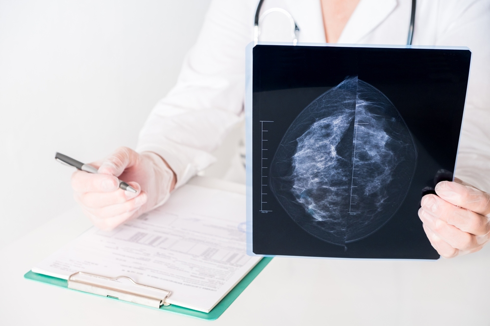
Contrast-enhanced mammography (CEM) has shown promise for women at higher risk of breast cancer, with follow-up screening showing greater specificity and accuracy.
Cancer detection rates remained consistent during repeat screening, while specificity improved compared with baseline examinations.
Specificity shows how accurately a test identifies people who do not have the disease, helping to reduce unnecessary follow-up procedures.

Researchers at Memorial Sloan Kettering Cancer Center analysed 6,911 contrast-enhanced mammography screens carried out among 2,756 women between 2015 and 2021.
Contrast-enhanced mammography, or CEM, combines standard mammography with an injected contrast agent that highlights areas of increased blood flow. Cancerous tumours often develop a greater blood supply.
The team compared 1,575 baseline screens with 3,336 incidence screens.
The researchers classified a screen as baseline if the woman had no previous CEM or had not undergone breast MRI in the previous three years.
Incidence screens were follow-up examinations carried out after previous screening.
After adjusting for the number of screens each woman received, the researchers found no statistically significant difference in cancer detection rates between the groups.
However, specificity reached 91.5 per cent during incidence screening, compared with 83.4 per cent for baseline screening.
Overall accuracy was also higher for incidence screening, at 91.4 per cent compared with 83.5 per cent.
“The ability of contrast-enhanced mammography to help detect cancer during prevalence screening in women at increased risk for breast cancer is maintained in subsequent incidence screens, with better specificity and accuracy,” the research team wrote.
Baseline screening detected 19 cancers per 1,000 examinations, compared with 11.6 per 1,000 incidence screens.
Sensitivity, which measures how well a test identifies people who have a disease, was 87.1 per cent for baseline screening and 86.1 per cent for incidence screening.
Contrast enhancement alone helped detect 18 of the 30 cancers found during baseline screening and 36 of the 62 found during incidence screening.
The researchers also identified 14 interval cancers across the examinations, with no evidence of a difference between the two groups.
An interval cancer is diagnosed after a screening result appears normal but before the next scheduled examination.
The retrospective study used previously collected clinical records rather than following participants in a newly designed trial.
The researchers described CEM as a reasonable screening tool for women with dense breasts.
Dense breasts contain more fibrous and glandular tissue, which can make cancer more difficult to detect with standard mammography.
CEM has previously shown greater sensitivity than ultrasound, digital mammography and digital breast tomosynthesis, according to the article.
Digital breast tomosynthesis takes several low-dose X-ray images from different angles to create a three-dimensional view of breast tissue.
The technique also takes less time and generally costs less than breast MRI, with previous research suggesting its performance is not inferior to MRI.
CEM is currently used for some screening purposes outside its approved indications.
The researchers said it could also help address health inequalities affecting women who face barriers to accessing MRI.
“In addition, women have reported a preference for CEM over MRI,” they wrote.
The researchers said a future article would provide full details about the interval cancers identified in the study.
An accompanying editorial said the technique could become a practical part of breast cancer screening for selected groups, although challenges remain around wider adoption.
“Whether this potential ultimately translates into widespread implementation will depend on future studies evaluating not only diagnostic performance, but also patient outcomes, health care utilisation, and real-world feasibility across diverse practice settings,” wrote Dr Vivianne Aguilera Freitas of the University of Toronto.
Insight
Experimental treatment significantly slows progression of fatal brain disease in women, study finds

Davunetide may significantly slow the progression of a fatal brain disease in women, according to a new analysis of clinical trial data.
The findings indicate that women and men with progressive supranuclear palsy (PSP) may respond differently to the experimental treatment.
Progressive supranuclear palsy, or PSP, is a rare and fatal neurodegenerative disease.
Researchers at Tel Aviv University led the analysis and said the results reinforce the need for sex-specific approaches to neurodegenerative diseases.
Neurodegenerative diseases are conditions in which nerve cells in the brain or nervous system gradually lose function and die.
The team reanalysed data from a 52-week international clinical trial involving more than 300 people with PSP.
The disease is caused by the abnormal accumulation of tau protein in the brain. Tau is a protein found in nerve cells that builds up abnormally in people with PSP.
There is currently no effective drug treatment for the disease.
The work was led by professor Illana Gozes of the Sagol School of Neuroscience and the Gray Faculty of Medical and Health Sciences at Tel Aviv University.
The research team included current and former students Dr Guy Shapira, Jason Blatt and Liri Guz, together with professor Noam Shomron.
The original clinical trial found that Davunetide was safe but ineffective.
However, the researchers separated female and male participants and re-examined the data using updated assessment measures recommended by the FDA.
Women treated with Davunetide experienced a significant slowing of disease progression, while no similar effect was observed in men.
The treatment helped preserve essential movement and functional abilities, including balance, fine motor skills and everyday tasks such as using cutlery, buttoning clothes and washing the face and hands.
Fine motor skills are the small, precise movements needed for tasks involving the fingers and hands.
Treated women also showed significant improvements in language ability, working memory and overall cognitive function.
Cognitive function covers mental abilities such as memory, attention, language and problem-solving.
The analysis also identified profound molecular differences between women and men.
The relationship between levels of pathological tau in cerebrospinal fluid and clinical symptoms was completely reversed between the sexes.
Cerebrospinal fluid is the clear liquid surrounding the brain and spinal cord. A biomarker is a measurable sign that can indicate disease activity.
For example, language abilities declined significantly as tau pathology increased in women, but not in men.
The researchers said this suggests the disease mechanisms may work differently in women and men, potentially explaining their different responses to treatment.
According to professor Gozes, overlooking biological differences between the sexes may hide a genuine treatment effect.
“Our data show that analysing women and men separately is not merely a statistical exercise, but an essential tool for developing more effective treatments for neurodegenerative brain diseases,” she said.
The researchers believe the findings provide a strong scientific basis for future clinical trials and treatment protocols designed from the outset to account for patients’ sex.
These trials could evaluate Davunetide as a targeted treatment for women with PSP.
They said the approach may also pave the way for more precise treatments for tau-related diseases, including Alzheimer’s disease and other neurodegenerative brain disorders.
The study was supported by ExoNavis Therapeutics, which is developing Davunetide for brain diseases under licence from Ramot, Tel Aviv University’s technology transfer company.


A preeclampsia study has found unusual cell activity in mothers and babies that could reveal new targets for treatment.
The condition affects 2 to 4 per cent of pregnancies worldwide and is a leading cause of maternal and foetal mortality.
There is currently no cure, and severe cases can put both the mother and baby at risk.
Scientists from UCL and University College London Hospitals found that stressed placental cells, poorly functioning blood vessels and an overactive immune response all contribute to the condition.
Preeclampsia causes high blood pressure during pregnancy. It can affect blood flow to the baby and cause symptoms such as swelling, headaches, blurred vision and pain under the ribs.
Without treatment, it can damage the mother’s health, slow the baby’s growth and, in severe cases, become life-threatening.
Previous research has focused only on the placenta, the organ that develops during pregnancy to support the baby’s growth, rather than the tissues around it.
The researchers said the findings could reveal new therapeutic targets, which are biological processes that future treatments could be designed to alter.
Senior author professor Sara Hillman, of the UCL EGA Institute for Women’s Health, said: “We studied individual cells from both the mother and the baby to see how their activity changes in healthy pregnancies compared with preeclampsia.
“This helped us to confirm some changes already suspected in the condition and also discover new ones.”
The team studied 20 pregnant women recruited at UCLH, including 10 with severe preeclampsia and 10 without the condition.
They used genomic testing to examine individual cells in the placenta and other tissues where cells from the developing baby and mother come into contact.
Genomic testing examines genetic information to help researchers understand how cells behave and the roles they may play.
The other tissues studied were the myometrium, the muscular layer of the womb, and the chorioamniotic membranes, which surround the baby during pregnancy.
The team compared cells from healthy pregnancies and those affected by preeclampsia at different gestational ages, meaning different stages of pregnancy.
They used technology that can read the genetic information of thousands of individual cells at the same time, allowing them to see what each cell was doing and where it was located in the tissue.
In preeclamptic pregnancies where babies were born prematurely, before 37 weeks, during the third trimester, placental cells showed signs of stress and low oxygen levels.
The cells also did not use energy in the normal way.
Some cells responsible for reshaping the mother’s blood vessels were not working properly, the researchers found, which may affect blood flow to the baby.
There were also signs of an overactive immune response in the placenta, nearby tissues and the mother’s blood.
The researchers said this response, together with other stress molecules released by the placenta, helps explain why preeclampsia affects the whole body and can become serious.
They hope the findings will help researchers find treatments for the condition and potentially save lives.
Co-lead author Dr Yara Sanchez Corrales, of the UCL Great Ormond Street Institute of Child Health, said: “These findings point to specific biological processes that could be targeted with treatments. Acting early in pregnancy, especially in more severe early-onset cases, could help improve outcomes and reduce the high risks associated with severe preeclampsia.
“We hope that our findings may set us on the path to reducing premature births and fatalities associated with preeclampsia.”
Co-lead author Mr Theodoros Xenakis, of the UCL Great Ormond Street Institute of Child Health, said: “Future studies may provide an even clearer picture of the biological changes linked to the disease by including more participants and using even more precise methods.”











 News2 weeks ago
New menopause drug approved for use by NHS in Scotland

 Hormonal health2 weeks ago
Stardust period tracker shares health data, study reveals

 Entrepreneur1 week ago
Zero Candida market set to reach over US$2 billion by 2030 – report

 Entrepreneur2 weeks ago
Women’s health draws record $1.55bn in equity as capital spreads beyond the mega-rounds

 Entrepreneur2 weeks ago
Onto Health acquires diagnostics software company Levy Health

 News6 days ago
From Hologic to Ark Surgical: Why Joseph LaBruzzo is betting on the future of women’s surgical innovation

 Mental health1 week ago
Nearly 60% of young women get health advice from influencers, research finds

 News2 weeks ago
Congress urged to invest over $20bn to close women’s health gap