


Diagnosis
Why microplastics in intimate care demand a scientific response

Dr Olivia Ahn, founder of FLUUS
The FemTech industry is rapidly innovating, but the conversation often neglects a fundamental issue: material safety.
While we focus on digital tracking and advanced fabrics, the tiny, ubiquitous fragments of plastic in our personal care products, microplastics, pose a significant, yet often silent, threat to women’s intimate and systemic health.

As a former doctor who moved into material science, my research focuses on the intersection of these two fields, particularly exploring the pathways through which microplastics from period and intimate care products enter the bloodstream.
The current evidence demands a radical shift in how founders and manufacturers approach product design.
The Ubiquity of the Problem
Microplastics, defined as plastic particles smaller than five millimeters, are no longer confined to remote oceans or deep-sea trenches.
Recent studies have demonstrated their presence in every part of the human body, including the bloodstream, lungs, placenta, and reproductive tissues.
For the FemTech and consumer health sectors, this raises profound questions. While we often focus on microplastics shed from synthetic clothing or food packaging, we must address direct exposure pathways.
Many single-use period pads and liners contain synthetic polymers, adhesives, and backsheets, which are all sources of microplastic shedding through friction and degradation.
Even when the outer layer is organic cotton, the hidden core materials can present a systemic risk.
The Intimate Absorption Pathway: A “Fast Track” to the Bloodstream
The primary concern with microplastics in intimate care lies in the unique vulnerability of the vaginal and vulval mucosa.
Unlike the skin on our arm, which is designed to be a tough, protective barrier (keratinized epithelium), the vaginal mucosa is highly specialised and much more permeable. It is a richly vascularised tissue meaning it contains a high concentration of tiny blood vessels just beneath the surface.
This structure allows for:
1. Rapid Systemic Uptake
The tissue acts almost like a sponge, allowing substances to be absorbed quickly and directly into the circulatory system. In medicine, this pathway is leveraged for rapid drug delivery, confirming its efficiency.
2. Bypassing First-Pass Metabolism
When substances are absorbed through the gut, they pass through the liver, which acts as a primary filter to detoxify or metabolize compounds.
When chemicals or microplastics enter via the vaginal mucosa, they can bypass this critical “first-pass” mechanism, potentially leading to higher systemic exposure levels of the contaminants.
When microplastics are present in a period pad, making prolonged, intimate contact with the mucosa, there is a clear and high-risk pathway for these particles to cross the tissue barrier and enter the bloodstream.
The Broader Implications for Women’s Health
The health impact of microplastics is complex and still emerging, but existing evidence raises serious red flags, particularly for gynaecological and reproductive health:
Inflammation and Oxidative Stress:
Microplastics are known to induce inflammation and oxidative stress in cell cultures and animal models.
Chronic, low-grade inflammation is a foundational mechanism for numerous diseases, including endometriosis, Polycystic Ovary Syndrome (PCOS), and cardiovascular issues.
Microplastics has not been linked to be causative to these conditions, but reducing any source of inflammatory burden is critical for overall health.
Hormonal Disruption:

Dr Olivia Ahn
Microplastics can act as carriers, or “Trojan horses,” for endocrine-disrupting chemicals like phthalates and BPA, which are added during plastic manufacturing.
When the microplastic enters the body, it releases this cocktail of EDCs, which mimic or interfere with natural hormones.
This disruption is directly linked to fertility challenges, impaired ovarian function, and altered reproductive development.
Reproductive Tissue Accumulation:
Recent studies are particularly alarming, detecting microplastics in human ovary follicular fluid, semen, and the placenta.
These findings suggest that microplastics are accumulating in the very tissues responsible for reproduction and fetal development, demanding immediate action to minimise exposure.
Setting a New Standard for Integrity
The challenge for founders and manufacturers is no longer merely to sell a product, but to assume full responsibility for its material science and its entire lifecycle.
We must move past the industry’s status quo where products are chemically complex and built to last centuries.
We must advocate for radical transparency in ingredient disclosure and invest in genuine, circular technology that eliminates these pollutants at the source.
The next generation of femtech must prioritise both the user’s health and the planet’s health equally
The Fluus Standard: Zero Microplastics, Zero Waster and Zero Compromise
This drive for scientific integrity is the foundation of Fluus.
We developed our proprietary Flushtec technology to prove that a 100 per cent microplastic-free, fully flushable period pad is not just an ideal, but a reality.
By eliminating plastic SAPs and traditional hot-melt acrylic adhesives, we deliver genuine confidence, ensuring the product fully disintegrates after use, leaving zero waste and zero microplastic residue behind.


Article produced in association with Spital Clinic
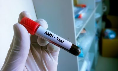
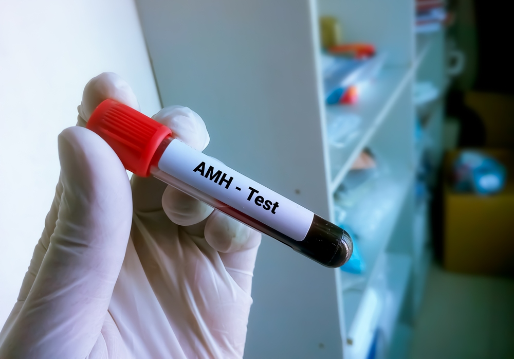
AMH has become one of the most-requested blood tests in private women’s health. The number it gives back is useful, but only when it is read in context.
AMH testing in the UK has gone mainstream over the past few years. Home-testing kits sell it as a snapshot of “your fertility”.
Private clinics include it in screening packages. On social media, individual AMH results are now routinely treated as a verdict on whether a woman will be able to have children.
That reading isn’t accurate. Anti-Müllerian Hormone (AMH) does carry useful information, but only inside a wider clinical picture.
Looked at on its own, it produces a lot of unnecessary anxiety, and often hides the questions that matter more.
What AMH measures
AMH is a hormone produced by the small follicles in the ovaries, the ones that haven’t yet been recruited for ovulation. Because these follicles are relatively stable across the menstrual cycle, the test can be done on any day, without needing to be timed to a period.
A higher AMH level tends to indicate a larger pool of these follicles. A lower level suggests the pool is smaller. That, broadly, is what the result shows.
The HFEA, the UK’s independent regulator of fertility treatment, describes AMH as an indicator of ovarian reserve, while making clear that fertility test results of this kind “are not guaranteed” as a predictor of fertility outcomes.
Put simply: AMH is a count of what is there. It says nothing about how well the body will use it, and it cannot predict if or when conception will happen.
Where AMH fits in a modern fertility assessment
In current UK private practice, AMH is rarely tested in isolation. A meaningful fertility assessment will pair it with a fuller hormone profile (FSH, LH, oestradiol, prolactin and thyroid function), along with markers such as Day 21 progesterone, vitamin D and rubella immunity where relevant.
This is the structure used in a trying-to-conceive screening, and there is a reason for it: each of these tests answers a different question that AMH on its own cannot.
It is this combination, not the AMH number on its own, that gives a clinician enough information to say anything meaningful about an individual’s reproductive picture.
Misconception 1: “A low AMH means natural pregnancy isn’t possible”
This is the misconception that causes the most distress, and it is consistently wrong.
Several large prospective studies of women in their 30s and 40s trying to conceive naturally have found that women whose biomarkers, including AMH, pointed to a diminished ovarian reserve were no less likely to conceive within twelve cycles than women with reassuring results.
That is why neither UK regulators nor national guidance treat AMH as a test that can predict natural fertility in women who have no known infertility issue.
The reason is simple. Natural conception only requires one good egg, released in a normal cycle, in the right window.
AMH doesn’t measure egg quality, and it doesn’t reveal whether ovulation is happening. A woman with low AMH may still ovulate every month with high-quality eggs.
A woman with high AMH (often the pattern seen in polycystic ovary syndrome) may not be ovulating regularly at all.
The NHS emphasises that age is the strongest single predictor of natural fertility. A 35-year-old with a low AMH and regular cycles is, on average, more likely to conceive naturally than a 40-year-old with a normal AMH and irregular ones.
If AMH comes back low for someone who is trying to conceive, the more useful question isn’t whether pregnancy is still possible (the answer is almost always yes), but whether there is reason to investigate the wider picture now rather than waiting twelve months.
Misconception 2: “A normal AMH means everything is fine”
The opposite assumption is just as risky.
AMH tells you about egg quantity. It does not tell you about:
- Egg quality, which is closely tied to age
- Whether ovulation is happening regularly
- Whether the fallopian tubes are open
- Whether there are structural issues such as fibroids, polyps, ovarian cysts or endometriosis
- Sperm parameters in a male partner
- Whether implantation will succeed
A reassuringly normal AMH at 38 still sits alongside age-related changes in egg quality. A slightly lower-than-average AMH at 28 may carry no real-world implications at all.
That is why no UK clinical body recommends AMH as a routine screening test for healthy women who have no fertility concerns. NICE’s fertility guideline, NG73, treats AMH as one component of a broader investigation, not as a verdict in itself.
Imaging is the natural counterpart to the blood test. A transvaginal pelvic ultrasound directly visualises the small follicles that produce AMH, the antral follicle count. It also picks up structural findings a blood test will never reveal, including ovarian cysts, fibroids, polycystic ovarian morphology, and abnormalities in the uterine cavity. A full ovarian reserve assessment normally includes both.
Where the AMH number actually matters
There are three settings in which AMH carries real, decision-relevant information.
Before IVF or egg freezing. AMH is one of the better predictors of how the ovaries are likely to respond to stimulation medication.
A higher AMH usually predicts more eggs collected per cycle, and a very low AMH may shape decisions about protocol or whether to bank cycles before treatment.
During a fertility investigation. If a couple has been trying for twelve months, or six months if the woman is over 35, AMH becomes part of a wider assessment that should also include ovarian ultrasound, a fuller hormone profile, semen analysis and an assessment of tubal patency.
As context for women planning ahead. Women who want to understand their reproductive options before they are ready to conceive (for example, ahead of a decision about egg freezing) can find AMH informative, provided it is interpreted alongside age, antral follicle count, and other markers, by a clinician who can place the number in context.
Reading the number properly
For anyone who has had an AMH test, three things make the result more useful:
- Pair it with age. A “normal” AMH at 25 means something very different from the same number at 38. Age is doing more work in the equation than the AMH value itself.
- Pair it with imaging. Ultrasound shows what is actually in the ovaries today, rather than relying on a single biochemical marker.
- Read it with a clinician. A number on a screen, with no context, no follow-up and no plan, is the worst way to use a test that, properly interpreted, can be very informative.
AMH is a useful tool. It just isn’t the headline it has often been turned into.
Disclaimer
This article is produced for informational purposes only and does not constitute medical advice, diagnosis or treatment. Clinical guidance referenced reflects published HFEA, NHS and NICE information available as at May 2026. Individual circumstances vary; readers are advised to consult a qualified healthcare professional before acting on any information in this article. This piece was produced in association with Spital Clinic, which provided background clinical information for editorial purposes. Hyperlinks to external sources are included for reference only and do not represent an endorsement of any product, service or organisation.


Female sex may not raise stroke risk across all atrial fibrillation (AF) patients, with higher risk mainly seen in women aged 75 and older, a study suggests.
Researchers said stroke prevention for women with the condition should be more personalised, especially for patients under 75.

Dr Amitabh C Pandey, director of cardiovascular translational research at Tulane University School of Medicine, said: “For years, female sex has been included as a risk factor along with other factors such as high blood pressure and diabetes, meaning women were more likely to be prescribed anticoagulants.
“Our study shows younger women may not have as much added stroke risk as previously thought, while older women, particularly those over 75, appear to have a higher risk that deserves close attention.”
The new Tulane University study challenges a long-standing assumption in heart care that being female automatically increases stroke risk for patients with atrial fibrillation.
Atrial fibrillation, often called AF, is a common heart rhythm disorder that causes the heart to beat irregularly.
It is associated with a higher risk of stroke and is often treated with anticoagulants, also known as blood thinners.
The study found that stroke risk did not increase equally across all female patients with AF.
Instead, researchers said being female may act more as a risk modifier, with increased stroke risk seen primarily among women aged 75 and older or those with a greater burden of other health conditions.
Clinicians often use a scoring system to decide whether people with AF should be prescribed blood thinners.
The system gives points for factors including age, heart failure, diabetes, previous stroke, vascular disease and high blood pressure.
Women also receive one point for sex alone.
Researchers said this can mean women with AF become eligible for blood thinners earlier or more often than men with otherwise similar risk profiles.
While blood thinners can help prevent clot-related strokes, they can also increase the risk of bruising, prolonged bleeding, gastrointestinal bleeding and other serious complications.
The researchers analysed approximately 950,000 patients with AF using TriNetX, a large anonymised electronic health record database.
They compared stroke outcomes between male and female patients across three age groups: younger than 65, 65 to 74, and 75 and older.
Male and female patients were matched based on age, other health problems and whether they had been prescribed anticoagulation medicine.
Among patients younger than 75, the study found no significant difference in one-year stroke risk between men and women.
However, among patients aged 75 and older, women had a modest but statistically significant increase in stroke risk compared with men.
In patients aged 75 and older with no additional risk factors beyond age, women had about one additional stroke per 629 patients compared with their male counterparts.
The findings support growing interest in a newer AF risk score, known as CHA2DS2-VA, which removes sex as a standalone risk factor.
However, researchers said more studies are needed and medical guidance remains inconsistent.
Han Feng, assistant professor at Tulane University School of Medicine, said: “This general approach came from women being underrepresented in AFib trials and studies comprising only about one-third of study populations.
“Our study shows not all women with AFib have the same risk profile, and these decisions should be individualised.
Pandey said: “These findings highlight the need for modern tools and approaches that can personalise risk profiles to individuals.
“The goal is not to undertreat patients who need stroke prevention, but to better identify who is most likely to benefit from anticoagulation and who may be exposed to unnecessary risk.”


 Menopause2 weeks ago
Perimenopause misinformation ‘putting women at risk’

 Hormonal health3 weeks ago
NHS urged to update website following renaming of PCOS

 Insight4 weeks ago
PCOS renamed after decade-long campaign to end ‘cyst’ misconception

 News1 week ago
Women still being failed when they reach menopause, experts say

 Entrepreneur2 weeks ago
Women’s Health Innovation Summit opens submissions for 2026 Innovation Showcase

 Diagnosis4 weeks ago
Artera receives FDA Clearance for breast cancer platform

 News2 weeks ago
Three menopause innovators shortlisted for Femtech World Award
 Fertility3 weeks ago
AI could transform ovarian care through personalisation, study finds