




Pregnancy
AI software designed to transform prenatal ultrasound receives FDA clearance


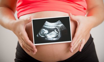
BrightHeart has announced it has received FDA clearance for its first artificial intelligence (AI) software, designed to transform prenatal ultrasound evaluations of the foetal heart.
Foetal heart ultrasounds exams are essential for detecting congenital heart defects (CHDs), which are the most common type of birth defect, affecting nearly 1 in 100 newborns.
However, misdetection or delayed diagnosis can lead to severe consequences, including missed opportunities for life-saving interventions. BrightHeart’s AI software enables clinicians to improve the detection of morphological abnormalities suggestive of CHDs, in resource-constrained practices.
“Foetal heart assessments are among the most technically demanding aspects of prenatal ultrasound,” said Cécile Dupont, BrightHeart CEO and a Partner at Sofinnova Partners’ medtech accelerator, MD Start.
“Our AI-powered solution not only assists clinicians in detecting signs of potential abnormalities earlier but also enhances their confidence in confirming normal findings, which is equally critical for the peace of mind of expectant families.”
BrightHeart’s technology is designed to integrate into existing workflows, reducing the burden on overworked sonographers and OB/GYNs.
“We’ve moved from concept to FDA clearance in record time, demonstrating the potential of our product and the agility and commitment of our team,” added Cécile Dupont.
With FDA clearance secured, BrightHeart has said it is poised for its U.S. commercial launch, driving efforts to scale operations and enter the market.
“Resource constraints and workforce shortages are critical challenges in prenatal care, and we believe BrightHeart is uniquely positioned to address them,” said Michael Butchko, Chairman of BrightHeart.
“In this next phase, we aim to deliver our transformative technology to clinicians and expectant families, making a measurable impact on prenatal care outcomes.”




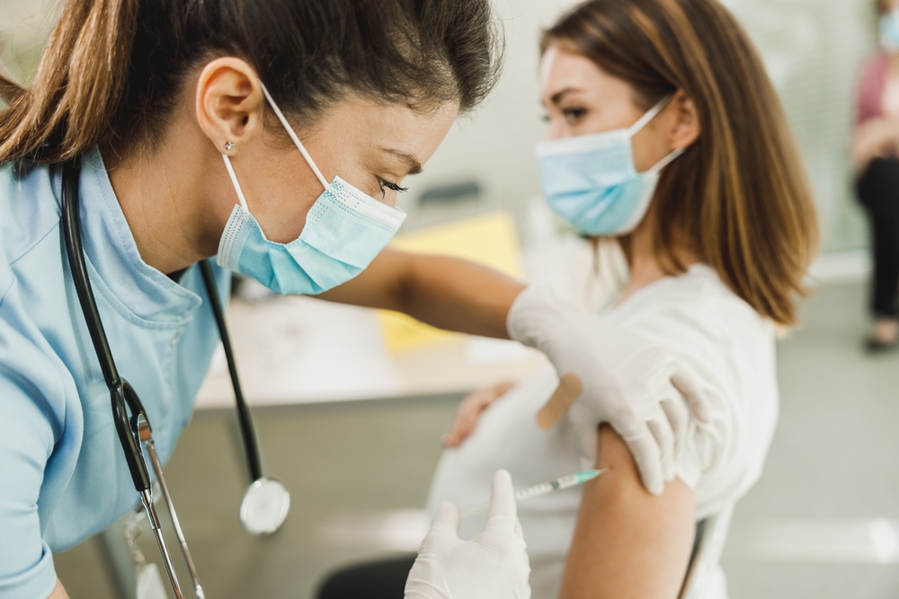
Pregnant women are among the first groups offered the NHS flu vaccine this winter, which has been updated to better match the subclade K strain.
Schoolchildren are also being prioritised and, like pregnant women, can receive the vaccine from September because they can catch and spread flu easily.
Protecting them also helps others, including younger siblings and grandparents.
Other eligible groups, including people aged 65 and over, will be able to receive a free NHS flu vaccine from October and should receive an invitation by phone or post.
Dr Amanda Doyle from NHS England said: “Last year’s flu season came early and was made worse by a new variant, which had a major impact for patients and put enormous pressure on urgent and emergency care services across the country.
“With the NHS experiencing its busiest summer on record, staff have started preparing for this winter earlier than ever – and with services already under significant pressure, it is vital that everyone offered the vaccine comes forward.”
Last winter’s flu season began more than a month earlier than usual as a new mutated strain circulated.
The strain was sometimes referred to as “Super flu”, although this is not a medical term and does not mean the virus is harder to treat. The name reflected that people had yet to build up immunity against it.
Flu vaccines are updated each year to better match the influenza viruses expected to circulate. These viruses continually mutate, so experts must predict how they are likely to change.
This winter’s vaccine includes changes intended to provide a better match for subclade K.
It remains unclear when flu will begin circulating widely this winter or how severe the season will be. Experts often look at flu activity in southern hemisphere countries such as Australia and New Zealand for indications of how the UK season could develop.
Their flu season peaks in July or August. Australia has recorded fewer cases than last year, while New Zealand has experienced high numbers.
Everyone is at risk of catching flu, regardless of age or how fit and healthy they are.
Doyle said: “Flu can really knock you for six, and for some children and pregnant women it can mean becoming seriously ill or ending up in hospital.”
The flu vaccine cannot give you flu. Instead, it helps the body know how to fight the virus.
Children aged two and over receive a nasal spray version, while adults receive an injection. One dose is needed ahead of winter.
Free NHS flu vaccines are available through pharmacies, GPs and schools. People who are not eligible for a free vaccination can pay around £10 to £20.
Schoolchildren and pregnant women can receive a free NHS flu vaccine from September onwards.
From October, free vaccination will also be available to children aged two or older who have not yet started school, people aged 65 and over, those with certain long-term health conditions, care home residents and carers of older or disabled people, including those receiving a carer’s allowance.
It will also be available to people living with someone who has a weakened immune system and those experiencing homelessness or staying in a homeless hostel or night shelter.
More adults will also be offered vaccination against respiratory syncytial virus (RSV), a virus that attacks the lungs, this winter.
From September, adults with certain pre-existing health conditions, such as chronic lung disease, will be invited for the RSV vaccine when they turn 65 rather than waiting until 75.



KOVE Medical has raised €1.7 million to support first-in-human studies of an implantable device designed to reduce preterm birth risk after foetal surgery.
The University of Zurich spin-off is developing a device designed to close defects left by surgical instruments following fetoscopy, a minimally invasive procedure performed inside the womb.
Fetoscopies can treat life-threatening conditions before birth but require surgical instruments to pass through the uterine wall and amniotic sac.
This can weaken the protective membrane, causing fluid leakage or rupture and often leading to preterm birth.
KOVE Medical said in a statement on LinkedIn: “This is a crucial step forward for our company, and we are incredibly grateful for the strong support and trust from our syndicate of visionary angel investors.”
KOVE Medical says its device closes the defect at the end of the procedure, stabilising the membrane and preventing complications. The aim is to make in-utero interventions safe enough to become a standard method of care.
The company plans to use the seed funding to reach first-in-human studies, a key milestone for a medical device seeking regulatory approval.
The CHF1.6m round was backed by a syndicate of angel investors.






 Hormonal health2 weeks ago
Major UK study could be a ‘game-changer’ for heavy periods and endometriosis
 Entrepreneur6 days ago
KOVE Medical raises €1.7 million to improve safety of foetal surgery

 Pregnancy2 weeks ago
One week left to apply: W Accelerate with Merck KGaA and M Ventures

 Pregnancy1 week ago
UK study aims to transform maternity care for high-risk pregnancies

 Menopause2 weeks ago
Quarter of women miss work due to menstual symptoms, survey finds

 Hormonal health2 weeks ago
PMDD after SSRIs or hormones: Why the brain may be the missing treatment target
 Diagnosis2 weeks ago
Ark Surgical secures further institutional backing to accelerate US expansion

 Menopause4 days ago
Menopause hormone treatment may ease brain fog, study suggests